![]()
Limb fracture and pinning of the
broken bone
Esther van Praag Ph.D.
|
|
MediRabbit.com is funded solely by the generosity of
donors. Every donation, no matter what the
size, is appreciated and will aid in the continuing research of medical care
and health of rabbits.
Thank
you |

Warning: this page
contains pictures that may be distressing for some persons.
Rabbits are prey animals that
must be able to flee and run rapidly. Their limbs are therefore powerful,
while their skeleton is light, comprising only 7 to 8% of the body weight. In
comparison, the skeleton of a cat makes up about 12 to 13% of the body weight.
Rabbits are more susceptible to fractures in their spine and limbs compared
to other animals.
Back injuries are frequently
observed at the lumbar level. Additional health problems, such as
osteoporosis and/or a calcium deficiency diet, can cause bones to become
brittle, increasing the risk of fractures. Such fractures can occur when a
rabbit panics, is improperly restrained or picked up, or is dropped. The
result is frequently paralysis of the lower limbs, accompanied by
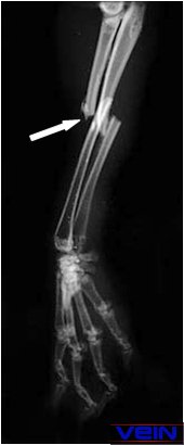
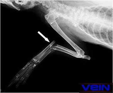
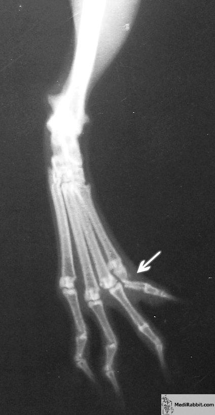
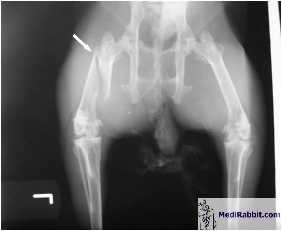
incontinence. Another risk for rabbits is a
fracture of the tibia. These injuries are frequently the result of improper
handling or caging practices, where a limb becomes entrapped in wire mesh
that is not the appropriate size. Fractures of limbs are frequently comminuted,
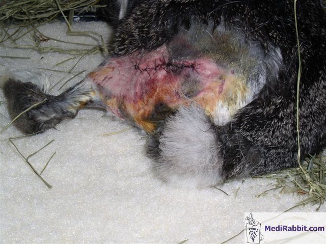
meaning they involve more than two broken pieces of bone. Fractures in the
lower part of limbs (below the knee) are often open due to the minimal
presence of soft tissue in this area. These wounds are often challenging to
treat and require patience to prevent further skin lacerations. The wound
must be sterilized, and the fractured bone pinned or splinted if the lower
part of the limb is affected. Osteomyelitis (bone infection) and/or gas
gangrene can rapidly develop, and must be treated promptly with
broad-spectrum antibiotics. It is also advisable to perform a bacterial
culture to determine which types of bacteria (aerobic or anaerobic) have
potentially contaminated the wound. Diagnosis
and first aid In the case of a rabbit with a
broken limb, emergency medical care is not required unless there are signs of
shock, bleeding, respiratory, or cardiovascular problems. It is recommended
that immediate pain medication be administered to ensure the rabbit's comfort
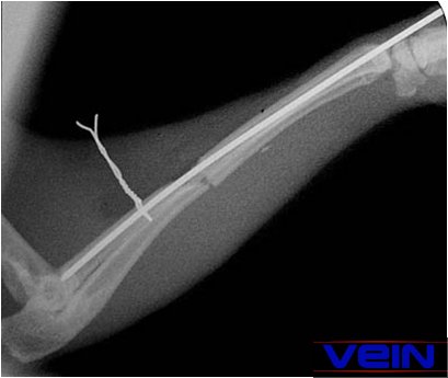
and tranquility. In
order to determine
the extent of the damage, the next steps for treatment, and the prognosis for
wound healing, it is necessary to perform X-rays and a complete physical and
neurological examination.
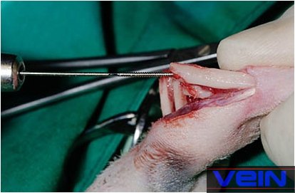
Use of intramedullary
fixation The intramedullary pinning
technique is a common procedure in small animals and is relatively
straightforward. This process necessitates patience and technical expertise,
as well as a solid understanding of biomechanics and bone healing. Intramedullary fixation is the
optimal treatment for rabbits, as the implant will help support the body's
weight. However, it should be noted that healing time may be delayed.
Steinmann pins, Kirschner wires, and/or cerclage wire are commonly used in
rabbits. Smooth intramedullary pins are generally preferred over partially or
fully threaded pins. In the case of dwarf or small rabbit breeds, the pin can
be substituted with a hypodermic needle. It is imperative that the pin or
needle penetrate 60 to 70% of the medullary center.
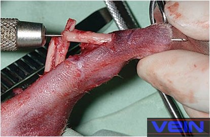
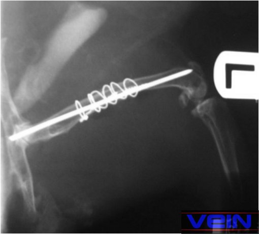
The Steinmann pin is typically
inserted manually or using a Jacobs chuck. The implant can be inserted either
at the fracture site (retrograde insertion) or at the end of the bone (normograde insertion), and it can be driven through the
fracture site. The protruding end of the pin is cut to prevent damage to the
skin, bacterial infection, or interference with the joint. However, if an
external skeleton fixation system is used, the pin should be left protruding.
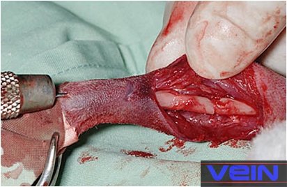
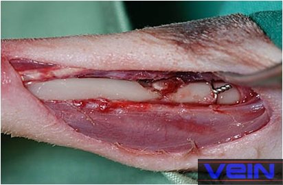
Open fracture repair techniques
necessitate appropriate sterile working conditions, accompanied by thorough
cleaning of the surgery site. Depending on the location of the
fracture, the appropriate treatment may include the use of padded bandages,
auto-adhesive bandages, or casts.
Post-surgical
treatment Post-surgical care involves regular examination of
the fracture site for signs of infection, pin or needle loosening, and
fracture healing. The removal of the pin is based on X-ray pictures.
Typically, this occurs approximately six weeks after the procedure. Rarely,
if a health complication affects the rabbit, the pin may be left in the bone.
Pain is a
signal that prompts the body to protect itself and reduce activity in the
affected area to minimize further injury. It is detrimental because prolonged
inactivity and spasmodic activity can result in weakness, loss of muscle
tone, and irreversible damage. Pain medication is therefore of the utmost
importance. A variety of effective analgesics are available to address
orthopedic pain: • Opioids: -
Butorphanol; -
Buprenorphine. • NSAIDs: -
Flunixin; -
Meloxicam; -
Aspirin. See “Analgesics drugs for
use in rabbits” for dosages. Furthermore, pain can lead to a reduction in
appetite and a decrease in water intake. It is therefore essential to ensure
that the rabbit is adequately hydrated. Acknowledgement I
would like to express my sincere gratitude to Sandy Minshull and Akira
Yamanouchi (Veterinary Exotic Information Network, http://vein.ne.jp/) for
their kind permission to use their pictures. I would also like to express my
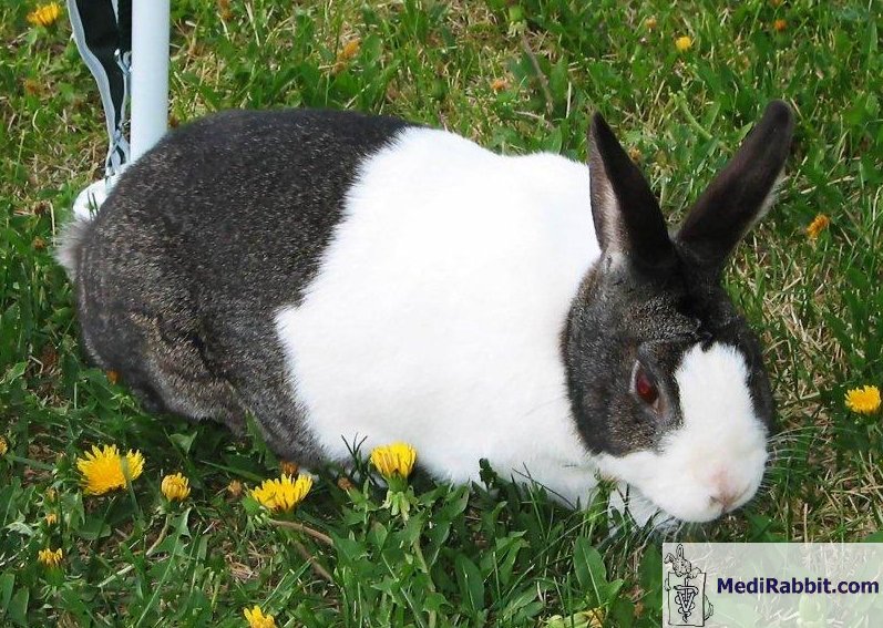
gratitude to Herman, who is 11 years old. Further
information Flecknell P. BSAVA Manual of Rabbit Medicine and
Surgery, UK: British Small Animal Veterinary Association; 2000. Hillyer E.V. and Quesenberry K.E. Ferrets,
Rabbits, and Rodents: Clinical Medicine and Surgery, New York: Saunders;
1997. Manning P.J., Ringler D.H., Newcomer C.E. The
Biology of the Laboratory Rabbit, New York: Academic Press; 1994. Richardson V., Rabbits: Health, Husbandry and
Disease. Blackwell
Science; 2000. |
|||||||||||||||||||

e-mail: info@medirabbit.com
