![]()
The liver, a delicate organ in
rabbits
Michel
Gruaz -
Esther van Praag, Ph.D.
|
|
MediRabbit.com is funded solely by the generosity of
donors. Every donation, no matter what the
size, is appreciated and will aid in the continuing research of medical care
and health of rabbits. Thank you |

|
The liver is a large organ that
occupies a significant portion of the rabbit's abdomen. Its surface is
homogeneous and red-brown in color. The liver performs both endocrine and
exocrine functions, including bile secretion. The liver of rabbits is divided
into two major lobes, right and left, separated by a median fissure. The
accessory lobes, the caudate lobe and the square lobe, are located between
these main lobes. The liver is maintained in place in the abdominal cavity by
ligaments connected to the diaphragm and to the dorsal wall of the abdomen.
The liver plays a central role in the synthesis of proteins, in the
metabolism of sugars, and the storage of nutrients. It is also involved in
the degradation of toxins and the treatment of organic waste produced by
cells of the body. It furthermore has exocrine function, with bile secretion.
This balance can be disrupted by various factors, including disease, the
presence of bacteria, parasites, or toxins in the diet. Liver disorders are
often underdiagnosed in rabbits because the clinical signs are non-specific
and lack clear characteristics. Single
and double gall bladder The gall
bladder is a hollow, pear-shaped organ that lies against the liver in the
caudal surface depression. It produces bile, a viscous yellow-greenish liquid
that is released into the small intestine via the bile duct. This secretion
facilitates the digestion of fatty foods. In rabbits, bile is produced
continuously and is stored in the gallbladder before its release into the
small intestine. Bile from rabbits is primarily composed of biliverdin, which
possesses potent antioxidant properties, as opposed to bilirubin. The
quantity of bile produced by this animal per day is substantial, at
approximately 250 milliliters. Based on weight, this is seven times the
amount produced by a dog.
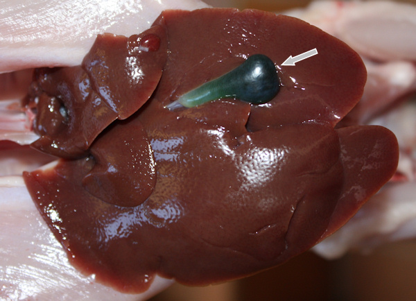
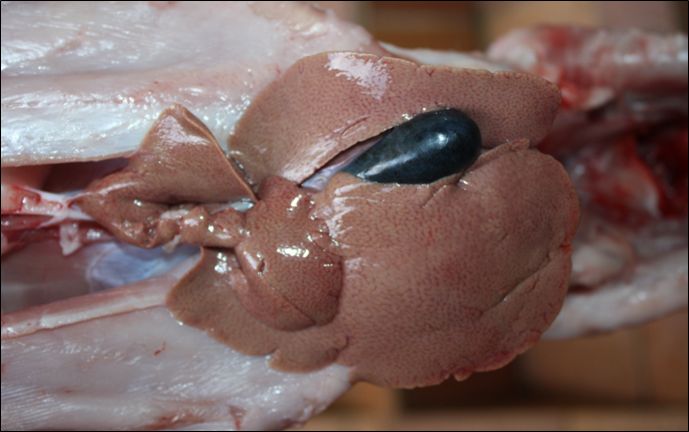
Normal
rabbit liver and gallbladder (arrow) The presence of a double
or bilobed gall bladder is an uncommon occurrence. The gallbladder
undergoes its initial development during the embryonic phase. The gallbladder
is subdivided, resulting in two complete vesicles of normal size and volume.
Their respective ducts transport bile to the intestine. These ducts have the
capacity to either merge halfway or to remain separate. The duplication of
the gallbladder appears to affect herbivores more than carnivores or humans.
The duplication of the gallbladder seems to affect herbivores more than
carnivores or man: 1 case out of 28 in cattle and 1 case out of 85 in sheep. Some
cases have been reported in rabbits as well. This congenital anomaly is
associated with various clinical manifestations, including pain and loss of
appetite. In other animals, a duplicate gallbladder is often associated with
biliary colic, acute inflammation of the gallbladder, or the presence of
gallstones.
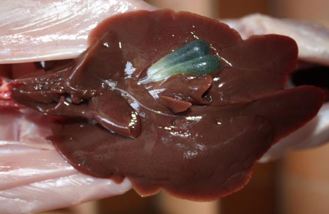
Michel
Gruaz Liver and double
gallbladder (pocket filled with green liquid - arrows) in a Harlequin rabbit
aged 3 months. Since weaning, this rabbit had regular digestive problems and
weighted only 2.3 kilos, while his littermates weighed 2.8 kg. Liver
lobe torsion The torsion of a lobe of the
liver is a sporadic (non-hereditary) event whose cause is not well clarified.
It is likely associated with an abnormal dilation of the stomach and
intestine following an intestinal obstruction. The ligaments that hold the
liver in place in the abdomen are distended and weakened, allowing the
twisting of a lobe. Other potential causes include external trauma, bacterial
or parasitic infection, or congenital absence of ligaments. A genetic
predisposition has been identified in the Madagascar lop rabbit population.
However, this remains to be verified, as the line of affected rabbits could
not be established. Gestation does not cause hepatic lobe torsion in rabbits
or other animals. Torsion of the caudate lobe is observed in 63% of
cases in rabbits. This lobe is attached to the liver on a very small surface,
facilitating its movement in the abdominal cavity or its torsion. This
results in impaired blood circulation, leading to atrophy of the lobe. The
lobe's consistency changes, becoming harder and acquiring a darker color. The
condition is painful and leads to decreased appetite, shock, and death after
a few days. In the event of a lobe rupture, the resulting hemorrhage can lead
to rapid death. A small percentage of rabbits have a positive outcome when
they receive supportive care consisting of oral food assistance with a
syringe, sterile subcutaneous fluids, analgesic, antimicrobial/anticoccidial,
and other intestinal motility drugs. (1 to 2 cc of olive oil has also been
found to be effective.).
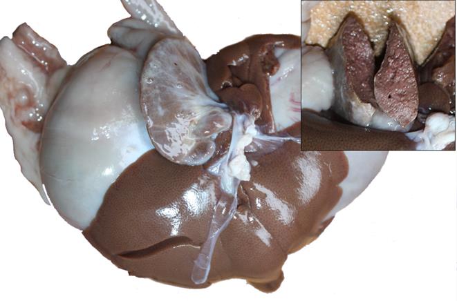
Michel
Gruaz Liver with torsion of
the small caudal lobe and content of the caudal lobe. Bacteria, coccidia and
parasites Bacteria have the potential to invade the liver,
leading to the formation of abscesses. The most prevalent bacteria identified
in such cases are Pasteurella sp. and Escherichia coli. In the
event of contact with birds or cattle, there is also a risk of infection from
salmonella, listeria, or Yersinia sp., which causes
pseudotuberculosis. As for the parasitic protozoa infecting the rabbit's
liver, they include those causing coccidiosis (Eimeria stiedae) and
toxoplasmosis. Coccidiosis is a prevalent disease in young animals. Their
digestive system is particularly sensitive to this parasite during the
transition from milk to vegetable food, which is more difficult to digest.
Indications of a potential infection may include a reduction in appetite,
diarrhea, the presence of mucus between hard stools, weight loss, and, in
severe cases, death. A swollen belly is often observed in young rabbits.
Older and healthy rabbits develop immunity against this parasite. A thorough
examination of the liver reveals the presence of yellow-colored nodules on
the surface. These nodules are difficult to differentiate from a bacterial
abscess.
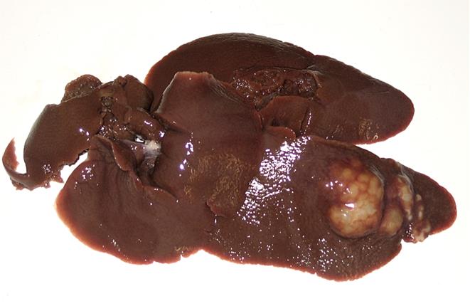
Michel
Gruaz These white structures
are difficult to differentiate between abscess and coccidia. Parasitic worms of the liver
are occasional in rabbits, typically resulting from the ingestion of food
contaminated by snails, dogs, or foxes. The rabbit does not host the adult
parasite, but rather an intermediate form. These include the liver fluke (Fasciola
hepatica), commonly found in grasslands, as well as the dog
tapeworm (Taenia sp.) and the fox worm (Echinococcus
multilocularis). The two latter form hydatid cysts containing the larvae
of the parasite. These cysts have the potential to occupy a certain volume in
the abdomen, thereby exerting pressure on the rabbit's liver. It is
challenging to detect these worms in live rabbits through clinical signs. Toxemia and liver lipidosis is a serious disorder that
occurs when there is a disruption of the energy metabolism in the late
gestational rabbit. Intrauterine growth of fetuses requires significant
energy. If this demand is not met with a high-calorie diet, the blood glucose
level of the doe drops rapidly. The adipose tissues are mobilized and begin
releasing fats into the bloodstream, which can lead to systemic poisoning.
The liver is no longer able to function properly and is unable to eliminate
ketone bodies. Other vital organs are also affected, including the kidneys.
The pH of the urine is reduced (5-6), its color is clear, and it contains
little sediments. However, it contains ketones and proteins. The intestinal
transit is decreased or even halted. It is not uncommon for affected rabbits
to experience diarrhea and/or a decrease in the amount of feces produced.
During autopsy, the organs, including the liver, thyroid, heart, kidneys, and
adrenal glands, are observed to appear pale. These organs may exhibit signs
of greasy infiltrations and necrotic foci. The stomach contains minimal food.
Hemorrhages are visible in both the placenta and the uterus. Another possible
consequence of a difficult gestation or lactation of a large litter is
hepatic lipidosis. The alternation of feed intake and refusal to eat can lead
to an accumulation of fats in the liver and kidneys of the doe. Their
functionality is altered. The rabbit's appetite diminishes, it exhibits
lethargy, and it experiences a reduction in body mass. Liver lipidosis can also be caused by dental
problems or a diet lacking in fiber. Rabbit obesity is characterized by an
abnormal accumulation of fat in liver cells linked to a diet that is too rich
in sugars (and not in lipids). Rather than metabolizing and eliminating
excess, the liver stores lipids in its tissues.
Michel
Gruaz Pale liver in a doe that
died from gestation toxemia. Toxins,
gas fumes and pesticides Various toxins have been shown
to affect the rabbit's liver and alter its functioning. Aflatoxin, a toxin
produced by molds, can contaminate cereals, affecting their quality and
safety. It is highly toxic to the liver and can result in a rapid onset of death.
There is a suspected link between plant toxins and neurological problems,
including head
down syndrome. The rabbit exhibits signs of lethargy, including a lowered
head and a hunched posture, often refusing to move or feed. The severity of
these symptoms varies from case to case. The prognosis for rabbits varies,
with some recovering after a few days, while others do not survive. Lead and
heavy metals have also been identified as factors contributing to liver
dysfunction. The use of wood products such as pine sawdust or cedar,
vermiculite, and other materials should be avoided in a cage or a hutch,
despite their pleasant smell. It has indeed been demonstrated that the
emanations of these products have toxic effects on the liver. The effects of Pesticides
on the nervous system may include hyperactivity, hypersensitivity, and/or
paralysis. These symptoms may be accompanied by breathing difficulties,
diarrhea, hypothermia, and/or nasal bleeding. Younger rabbits are more
sensitive and affected than adults. The general state of health and nutrition
of the animal is also a contributing factor. Treatment is essentially
supportive, involving the administration of assisted syringed food and fluids
to prevent dehydration. The autopsy of intoxicated rabbits reveals that the
liver exhibits a pale coloration, accompanied by cellular hypertrophy and
fatty degeneration. These disorders frequently occur before the animal enters
a coma and dies.
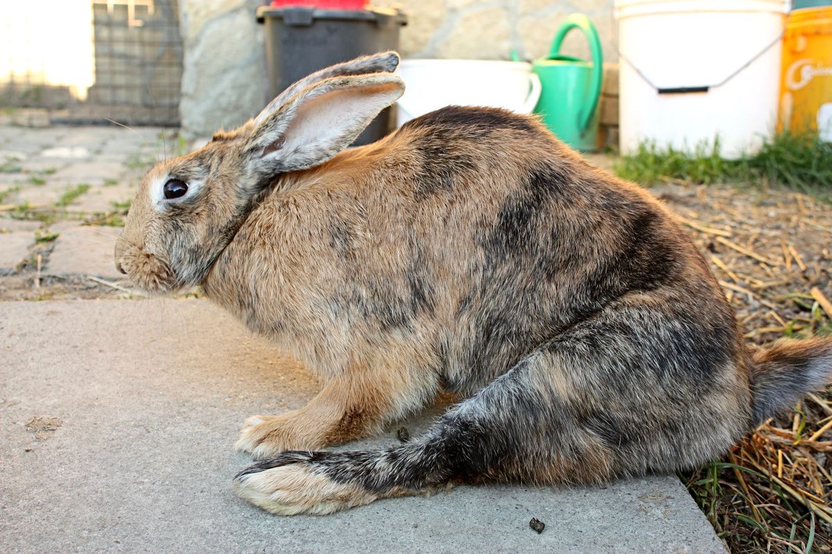
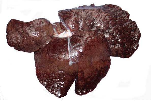
Typical
hunched position of a female rabbit, with paralysis of the hind-limbs, after
a pesticide intoxication.
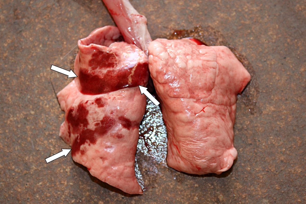
Lungs
of the above rabbit, showing multifocal hemorrhagic regions (arrows) in the
lungs. Deformation of the spinal cord A less common
cause of liver disease is bone
deformity of the spine, such as lordosis have been observed in rabbits,
though not consistently. The inward curvature of the spine exerts pressure on
the abdominal mass and compresses the blood vessels that drain blood from the
heart to the organs and vice versa. Liver and kidneys are primarily impacted.
In the liver, blood flow from the inferior vena cava is restricted, resulting
in reduced hepatic activity. The secretion of bile into the intestine is
decreased, which leads to a poor digestion of the ingested food. Consequently,
toxins accumulate in the liver, causing it to appear congested, dark red,
slightly larger than normal, and rounded with rough edges. Passive congestion
of the liver may be a secondary complication of heart failure.
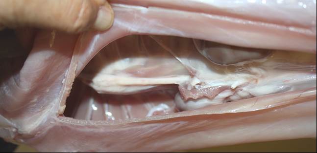
Michel
Gruaz The
following incidental findings were made during the necropsy: lordosis of the
spine (see arrow) and congested liver in a young Champagne-silver rabbit. The
spinal deformation is accompanied by liver congestion, which is a possible
consequence of compressed blood vessels from the heart to this organ. Finally, the rabbit's liver can
develop cancerous or bile duct tumors. Liver diseases are thus prevalent
among rabbits and often difficult to identify. Indeed, signs are not very
indicative of the presence of a disease. Additionally, rabbits tend to hide
any signs of illness. It is important to note the following observations:
droppings, their smell and shape, the presence of mucus, and the amount of
water drunk. In addition, veterinary tests must be performed to establish a
more precise diagnosis of liver disease. Further informationCere N, Humbert JF, Licois D, Corvione M,
Afanassieff M, Chanteloup N. A new approach for the identification and the
diagnosis of Eimeria media parasite of the rabbit. Exp Parasitol.
1996; 82(2):132-8. Cox JC, Edmonds JW, Shepherd RC. Toxoplasmosis and the wild rabbit Oryctolagus cuniculus in Victoria, Australia with suggested mechanisms for dissemination of oocysts. J Hyg (Lond). 1981;87(2):331-7. Coudert P., Licois D., Drouet-Viard F., Provôt F. 2000. "Coccidiosis". In: Rosell J.M. (ed), (Enfermedades del conejo), vol.II, chapter XVI, pp 219-234, Mundi-Prensa Libros, Madrid, Spain. Dubey JP, Passos LM, Rajendran C, Ferreira LR, Gennari SM, Su C. Isolation of viable Toxoplasma gondii from feral guinea fowl (Numida meleagris) and domestic rabbits (Oryctolagus cuniculus) from Brazil. J Parasitol. 2011;97(5):842-5. Grabarczyk M, Kopeć-Szlezak J, Szczepańska I, Woźniak J, Podstawka U. The effect of gamma-hexachlorocyclohexane (lindane) on blood cells, kidney and liver tissues in rabbits. Haematologia (Budap). 1990;23(3):171-9. Gustafsson K, Uggla A, Järplid B. Toxoplasma gondii infection in the mountain hare (Lepus timidus) and domestic rabbit (Oryctolagus cuniculus). I. Pathology. J Comp Pathol. 1997a;117(4):351-60. Kopeć-Szlezak J, Góralczyk
K, Woźniak J. Changes in serum and internal organs during increased
accumulation of gamma-hexachlorocyclohexane in adipose tissue of rabbits. Mater Med Pol. 1989
Oct-Dec;21(4):286-91. Licois D, Coudert P, Bahagia S, Rossi GL.
Endogenous development of Eimeria intestinalis in rabbits. J
Parasitol. 1992; 78(6):1041-8. Manning et al. The biology of the laboratory
rabbit. 2nd ed. London, UK, 1994. Milot L, Partensky C, Scoazec JY, Valette PJ,
Pilleul F. Double gallbladder diagnosed on contrast-enhanced MR
cholangiography with mangafodipir trisodium. AJR Am J
Roentgenol. 2005;184(3 Suppl):S88-90. Moores AL, Gregory SP. Duplex gall bladder
associated with choledocholithiasis, cholecystitis, gall bladder rupture and
septic peritonitis in a cat. J Small Anim Pract. 2007;48(7):404-9. Palmer AK, Bottomley AM, Worden AN, Frohberg
H, Bauer A. Effect of lindane on pregnancy in the rabbit and rat. Toxicology.
1978 Mar;9(3):239-47. Pakandl M, Drouet-Viard F, Coudert P. How do
sporozoites of rabbit Eimeria species reach their target cells? C R
Acad Sci III. 1995; 318(12):1213-7. Stingl H. Vesica fellea duplex and vesica
fellea divisa in rabbits (Oryctolagus cuniculus). Berl Munch
Tierarztl Wochenschr. 1971;84(21):420-2.
Stolkind E. Double gall-bladder
report of a case and review of 38 cases. British Journal of Surgery
Vol 27 (108), 1940:
760–766.
|
e-mail: info@medirabbit.com
