![]()
Anatomy of the tongue in rabbits
Esther van Praag, Ph.D.
|
|
MediRabbit.com is
funded solely by the generosity of donors. Every
donation, no matter what the size, is appreciated and will aid in the continuing
research of medical care and health of rabbits. Thank you |

Warning: this file contains pictures
that may be distressing to some persons
|
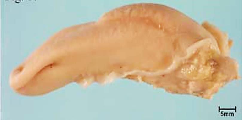
As with other mammals, the hyoid bone anchors the
rabbit tongue into the floor of the mouth. From this point, the elongated,
narrow tongue extends upward and forward. At six months of age, the total
length of the tongue is 65 millimeters, with the apex measuring 16
millimeters and the body measuring 37 millimeters. The width of the tongue
varies between 15 and 17 millimeters, depending on the region. The
protuberance of the posterior part of the tongue (torus linguae) is
fully developed. The median sulcus, which divides the tongue into symmetrical
halves, extends from the apex to the body of the tongue and ends in front of
the torus. On the ventral side of the tongue, a medial membrane (frenulum
linguae) connects the middle line of the tongue to the floor of the mouth,
thus limiting its movement. Extrinsic muscles restrict the movement of the
tongue and give it its characteristic convex form. These muscles include the
hyoglossus depressor muscle, the basioglossis
muscle, the ceratoglossus muscle, and the chondroglossus muscle, as well as the genioglossus and
the styloglossus muscles. The tongue's shape is highly adaptable and undergoes
changes during the processes of mastication and the retraction of ingested
material toward the rear of the oral cavity. This dynamic range of motion is
facilitated by a series of intrinsic muscles (lingualis proprius) located within the tongue's body.: - in the dorsal
part: longitudinal and superficial muscle fibers; - In the
central part: perpendicular and transverse muscle fibers; - In the
ventral part: longitudinal and deep muscle fibers.
As Cortopassi
and Muhl (1990) noted during videofluorographic
studies of the tongue during mastication: In the lateral view, the forepart
of the tongue moves down and forward during the opening stroke, whereas the
intermolar eminence moves up and forward to appose
the palate. During the closing stroke, as the tip of the tongue moves up and
back, the intermolar eminence lowers from the palate and retracts. During the
power stroke, the forepart of the tongue is at its most elevated and retruded position, while the intermolar eminence is at
its lowest and most retruded position. The dorso-ventral view demonstrated that the tongue and
mandible exhibit synchronous movement during lateral movement. The intermolar
eminence narrows during the power stroke, potentially twisting to position or
retain food between the teeth. Throughout the chewing cycle, the tongue
undergoes an anterior-to-posterior undulating movement. As the intermolar
eminence elevates to oppose the palate during the opening stroke, it may
replace the bolus on the teeth on the chewing side. The intermolar eminence
also appears to be twisting during the closing and power strokes to place or
maintain food on the teeth." This allows the ingested food to be pushed
backwards from the front-diastemal region to the
cheek teeth (premolars and molars), where food is placed on the mandibular
cheek teeth, and then chewed into small pieces. From this point, the ingesta
is directed towards the posterior region of the oral cavity. Swallowing is a
complex process that involves the coordinated action of the tongue, the soft
palate, and the pharynx muscles.
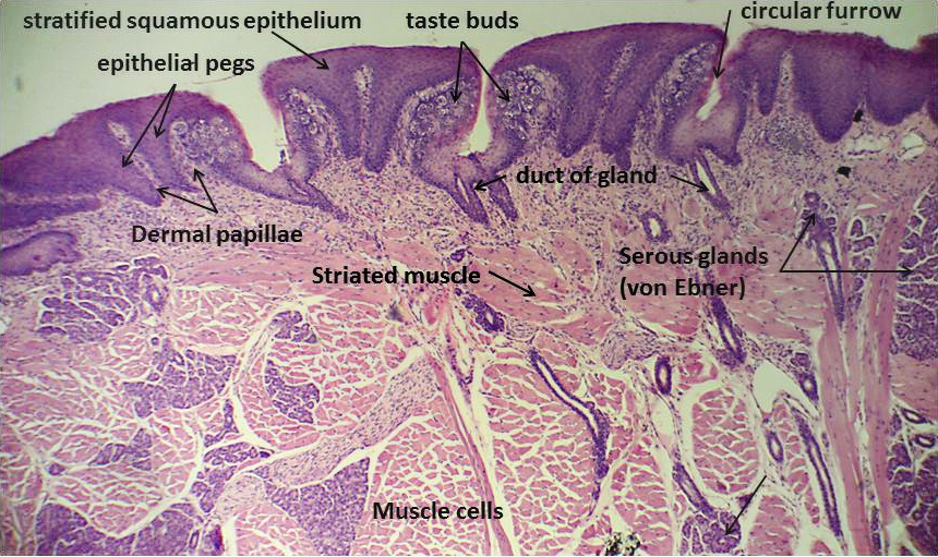
The dorsal
part of the tongue is divided into posterior smooth and hard portions, and
anterior smooth and rough portions. All possess extensions in the mucus
membrane that form the taste buds. Based on their location on the tongue,
different types of papillae can be distinguished: - Filiform
papillae – the numerous elongated conical papillae are typically observed on
the dorsal, softer anterior end of the tongue; - Fungiform
papillae – mushroom shaped projection found along the rostral margin of the
tongue. These cells contain taste buds, which are responsible for detecting
flavor. - Vallate
papillae – two dome shaped papillae are set symmetrically in the mucus
membrane of the apex and the body of the tongue and on the side of the torus.
Vallate papillae have been found to contain both taste buds and lymph nodes. - Foliata papillae –
these are well-developed in Lagomorphs and domestic rabbits. These papillae
are located in about 20 ridges on the posterior lateral sides of the tongue.
According to Engelman (1872), there are as many as 7,440 taste buds in the
circular furrows that line the walls of the papillae. There are open pores in
the cleft. Taste buds are connected to sensory nerve fibers,
which convey the sensory information to the brain. Further
Information Cortopassi D,
Muhl ZF. Videofluorographic analysis of tongue
movement in the rabbit (Oryctolagus cuniculus). J Morphol. 1990
May;204(2):139-46. Engelmann Th.W. The Organs of Taste. Strieker's Manual of
Histology,' New York, 1872. Kulawik M, Godynicki
S. Fungiform papillae of the tongue in the rabbit (Oryctolagus cuniculus).
Pol J Vet Sci. 2007a;10(1):25-7. Kulawik M, Godynicki
S. Vallate papillae in the domestic rabbit (Oryctolagus cuniculus f.
domestica). Pol J Vet Sci. 2007b;10(1):47-50. Kulawik M, Szymon Godynicki S.
Development of the tongue in the rabbit (Oryctolagus cuniculus f.
domestica) and the order of formation of lingual papillae in pre- and
postnatal life 1. Acta Sci. Pol., Medicina Veterinaria
8(4) 2009, 15-26. Ojima, K.;
Hosaka, M. & Suzuki, Y. Functional and positional difference and
classification of the fungiform papillae on the rabbit tongue seen in
microvascular cast specimens by means of scanning electron microscope. Ann.
Anat., 182(6):521-4,2000. Nonaka, K.; Zheng, J. H. &
Kobayashi, K. Comparative morphological study on the lingual papillae and
their connective tissue cores in rabbits. Okajimas
Folia Anat. Jpn., 85(2):57-66,2008. |
