![]()
Facial abscesses
Michel Gruaz, with Esther van Praag, Ph.D.
|
|
MediRabbit.com is
funded solely by the generosity of donors. Every
donation, no matter what the size, is appreciated and will aid in the
continuing research of medical care and health of rabbits. Thank you |

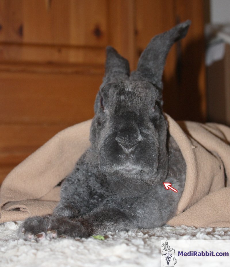
Warning: this page may contain pictures that may be distressing for some persons.
|
In the past, when a
rabbit developed a large abscess on its cheek, it was believed to be caused
by bovine hypodermosis, also known as 'warbles', which are caused by the
larvae of the ox warble fly (Hypoderma bovis). Lumps (sterile
abscesses) on the back of cattle or deer contain young warbles/larvae.
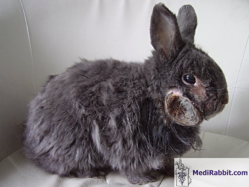
However, the presence of a facial abscess should be taken seriously. The main
causes are dental problems and bacterial infections such as staphylococcosis
and pasteurellosis. An abscess is a
pocket of fluid and pus resulting from an attack by pyogenic organisms
(bacteria that produce pus), followed by cell destruction. The contents
consist of pus, dead phagocytic white blood cells, necrotic cells, and dead
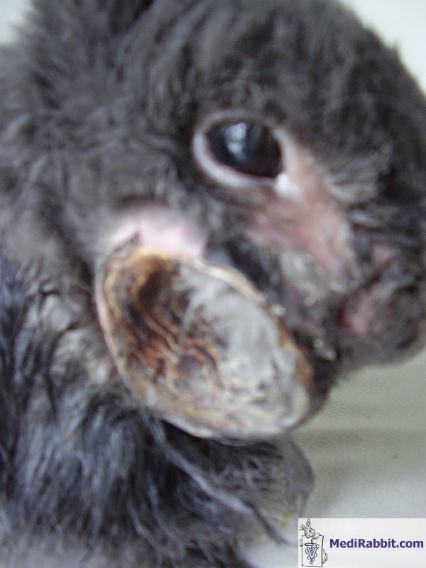
or living bacteria. The pocket grows in size as the amount of pus increases.
During the encapsulation phase, the abscess becomes isolated from the
surrounding tissues and blood circulation. If left untreated, the abscess can
burst either internally or on the skin's surface. This releases bacteria and
their toxins into the blood, which can be life-threatening and difficult to
treat in rabbits (known as septicaemia). Many rabbits
suffering from abscesses have a history of pasteurellosis or infections
caused by other bacteria, e.g. Streptococcus sp., Pseudomonas sp. and/or
fusiform – spindle shaped bacteria. The development of jaw abscesses may have
a genetic origin (malocclusion, abnormal elongation of a molar), but can also
relate to apical (tooth root) problems due to dental trauma (fracture) or to
the presence of a foreign body such as a piece of hay stuck between molars.
Bacteria have the capacity to penetrate the space between the gum tissue and
the tooth, ultimately reaching the dental root. Research has shown that Staphylococcus
sp. or Pasteurella sp. bacteria are often isolated, but it is
important to consider that Fusobacterium sp., Actinomyces sp.
or Streptococcus sp. (which can infect human teeth) may also be the
cause. There are no
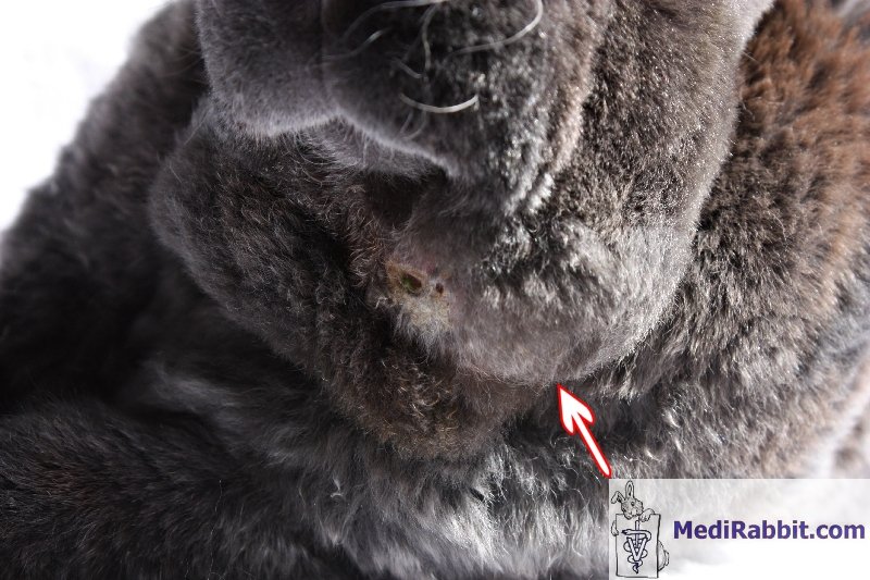
specific clinical signs that indicate the presence of an abscess. In the
initial stage, it is possible to feel a lump or a bump along the mandibular
bone. Abscesses can present as hard lumps or soft masses that are movable. It
appears that their presence does not cause pain in rabbits, in contrast to
the observations made in other animals. Abscesses tend to grow rapidly, with
their size doubling within a few days.
This stage is often
overlooked, as the rabbit continues to eat normally. Then, over time, an
unusual limp becomes apparent, accompanied by a reduced appetite, increased
drinking and, on occasion, fever.
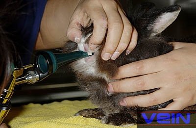
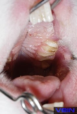
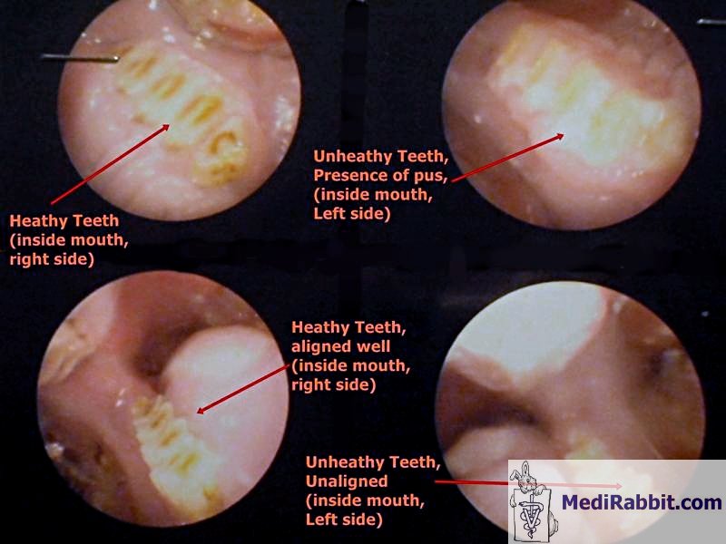
Identification of the problem - diagnosis
If
the visual examination and palpation of the head indicate the presence of a
maxillary or mandibular abscess, the oral cavity of the rabbit must be
carefully examined. This procedure is best carried out on a sedated or anaesthetized
rabbit.
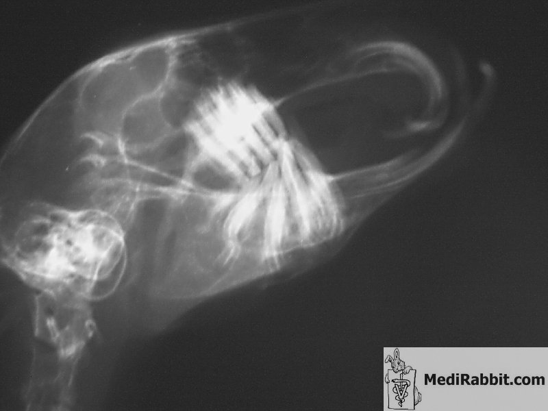
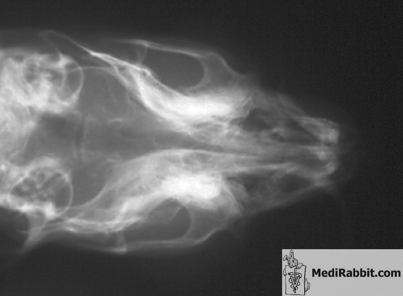
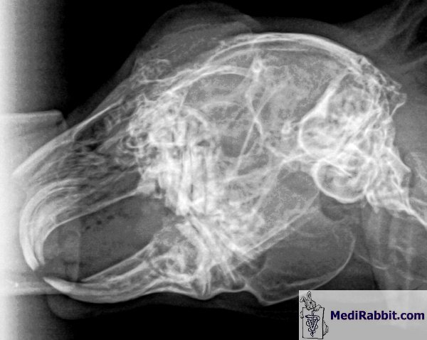
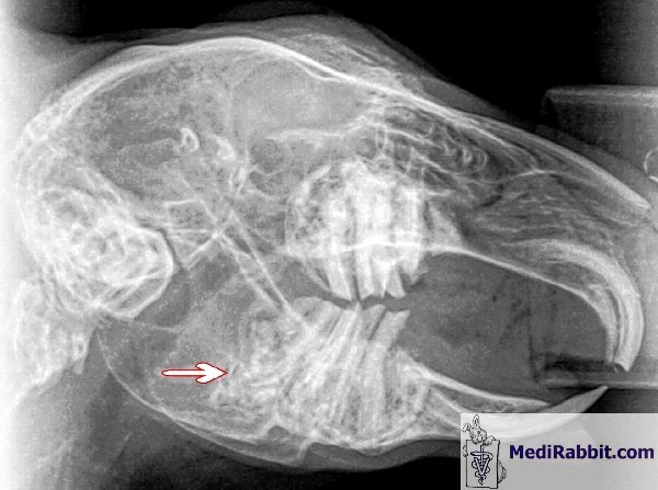
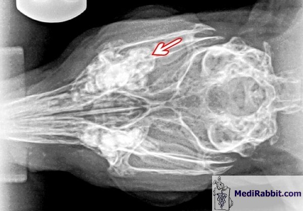
The anaesthetized
rabbit is immobilized to prevent movements that could compromise the clarity
of the images. Ideally, radiographs should be taken from different angles
(ventrodorsal, lateral et oblique). It is essential that images are of the
highest quality and resolution where possible. To provide information
regarding the dental problem, including but not limited to bone deformation,
root-related problems, the presence of abscesses, and the spread of infection
to the jaw bone. Computed tomography (CT) scanning is an invaluable tool
allowing precise identification of dental issues and assessment of their
severity. It facilitates the evaluation of prognosis and prediction of
treatment outcomes, contributing to informed treatment decisions.
Infection of the
jawbone is a risk. A sample can be aspirated in a syringe with a needle of
smaller gauge and sent to a specialized laboratory. It is unfortunate that in
some cases, the diagnosis may be incorrect. For example, the diagnosis may be
osteosarcoma (a tumour of the bone, which is rare in rabbits) instead of a
dental abscess with bone infection (osteomyelitis). Please be aware that this
error has the potential to have severe consequences, including delaying
appropriate treatment and administration of antibiotics. Treatment
The treatment of
facial abscesses is challenging and protracted, necessitating the owner's
cooperation and dedication to post-surgical care. Recurrence is frequent. If the lump can be
felt, but is small, an antibiotic treatment can be attempted. In one case,
the rabbit had lost a tooth, and the resulting cavity was filled with pus.
The combination of daily marbofloxacin and weekly injection of long-acting
penicillin successfully resolved the issue. If the rabbit is sensitive to
penicillin, this antibiotic can be replaced by metronidazole, for example.
However, the use of systemic antibiotics is not always effective. A more
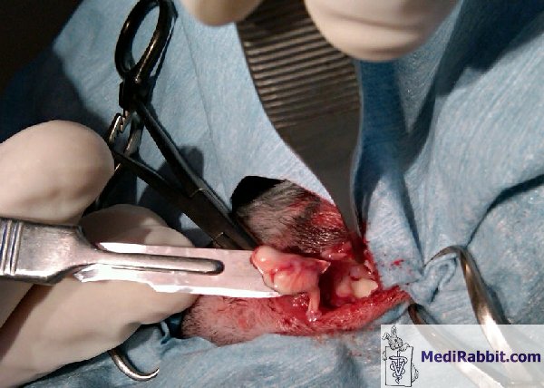
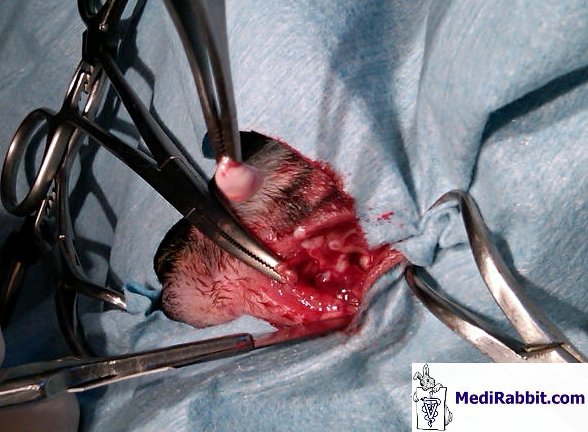
aggressive approach is therefore required. The safest approach is to excise the
abscess capsule and surrounding infected and/or necrotic tissues completely.
During the procedure, it is imperative to ensure that all fibrous channels
leading to abscess cavities located deeper in the tissue are removed. If the
pockets cannot be removed, they should be flushed with an antiseptic solution
(chlorhexidine or povidone iodine) via a catheter. The debrided cavity can be
filled with products impregnated with antibiotics. An alternative option
would be to place a drain to facilitate post-surgical care and promote
healing.
If
surgical excision is not feasible, the abscess pocket should be meticulously
debrided. It is essential to ensure that all traces of pus, tooth or bone
fragments, or necrotic tissue, are completely removed to promote optimal
healing. The pocket can be filled with PMMA beads impregnated with
antibiotics, cellulose-based sponges or calcium hydroxide, before the
incision is sutured. Details about fillings can be found here: Skin abscess in rabbits. Another option is
to leave the wound open by suturing the edges of the incision to the skin
(marsupialisation). This facilitates straightforward daily care, such as
flushing with chlohexiderme or povidone iodine, or filling with products that
dry out the cavity (e.g., dextrose, medical honey, Manuka honey). During the
healing process, the cavity will gradually be filled with scar tissue, thus
healing. In the case of
osteomyelitis, the extent of the infection in the jaw bone must be assessed.
If the infection spreads to several molars, the prognosis is guarded, and the
option of humanely euthanizing the rabbit should be considered. Treatment
involves the administration of systemic antibiotics that penetrate the bone,
selected based on the results of bacterial culture and antibiotic sensitivity
tests. The choice of antibiotics that are safe to use in rabbits is limited.
The treatment must be both aggressive and long-term, with a duration of
between four and six weeks. If the infection is not reduced by antibiotics,
or if bone has been destroyed, surgical debridement should be considered. Following the
procedure and throughout the recovery period, it is essential to administer
appropriate analgesics to the rabbit. There are a number of products on the
market which have healing properties, including creams and gels containing
Echinacea or HEALx Soother Plus. It is important to
note that abscesses can be challenging to treat and healing is not always
guaranteed. It is therefore recommended that post-surgical follow-ups are
carried out.. Acknowledgement
I would like to express my sincere
gratitude to Caroline Charland (www.BunnyBunch.org),
to Michel Gruaz (Suisse), to Debbie Hanson (USA) and her rabbit Stella, to
Dr. Cheryl Morales (Prestonwood Animal Clinic,
Houston, TX, US), to Bonnie Salt (USA), to Tal Saarony (USA) and her rabbit
Motek, to Dr. Gil Stanzione (Dakota
Veterinary Clinic, White Plains, NY, USA), to Jen Smuck (USA), and to
Akira Yamanouchi (Veterinary
Exotic Information Network, Japan) for kindly granting us permission to
use their pictures. Further
information
Capello V. Case Report: Use of
HEALx Soother Plus in Postoperative Treatment of a Dental-related Abscess in
a Pet Rabbit. Capello V, Gracis M, Lennox A.
Rabbit and Rodent Dentistry Handbook. Lake Worth - FL, USA: Zoological
Education Network; 2005. Harcourt-Brown F. Textbook of
Rabbit Medicine. Oxford, UK: Butterworth-Heinemann; 2001. Meredith A, Flecknell P. BSAVA
Manual of Rabbit Medicine and Surgery. Cheltenham, UK: British Small Animal
Veterinary Association; 2006. Quesenberry KE, Carpenter J.
Ferrets, Rabbits, and Rodents. St-Louis-MO, USA: Saunders; 2004. Van Praag E, Maurer A, Saarony T. Skin Diseases of Rabbits. Geneva, CH: MediRabbit.com; 2010. |
||||||||||||||||||||||||||||||||||||