![]()
Dental dysplasia: pseudo-odontoma (elodontoma) in rabbits
Esther van Praag, Ph.D.
|
|
MediRabbit.com is funded solely by the generosity of
donors. Every donation, no matter what the
size, is appreciated and will aid in the continuing research of medical care
and health of rabbits.
Thank
you |

Warning: this page may contain pictures that may be distressing for some persons.
|
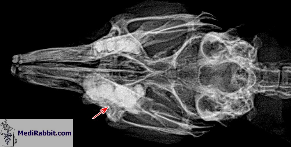
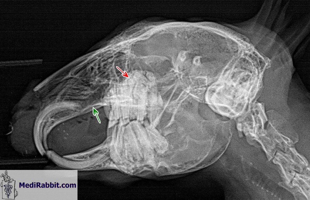
The growth of benign masses, such as
pseudo-odontomas, is rarely observed in rabbits. This dysplastic disease
involves the constant deposition of odontogenic tissue — including dental
pulp, mesenchymal cells, enamel, dentin, and cementum — around the roots of
the maxillary incisors or cheek teeth. The dysplasia is usually present on
both jaws. Etiology
The development of
pseudo-odontoma is not well understood. While it may be related to ageing, it
is more likely to be caused by an inflammatory process at the root of rabbit
teeth or osteoporosis. When the roots of incisors are affected, this may be
due to traumatic damage caused by chewing on cage bars, for example. Further
causes in rodents include viral agents or a nutrient- or vitamin-deficient
diet. Indeed, the disease has been observed in rats fed a vitamin A-deficient
diet. Clinical signs and
diagnosis
The early stages of
the disease are asymptomatic and there are no visible changes to the
appearance of the affected teeth. Therefore, it can only be recognized
through X-rays of the skull. At a later stage, the eruption of teeth is
impaired and modifications occur in the dental pulp canal. Chewing may become
difficult. Further clinical signs include anorexia, difficulty swallowing,
gastrointestinal problems, and reluctance to exercise due to dyspnea. Changes to the roots
of the maxillary incisor and cheek teeth, and deformation of the maxillary
bone, can lead to compression of the nasolacrimal duct and result in overflow
of tears (epiphora). Secondary upper respiratory difficulties are uncommon
unless the space-occupying mass encroaches upon the nasal cavity and airways.
Worsening obstruction of the air passage is characterised
by shortness of breath and inspiratory paroxysmal respiration ('reverse
sneezing'). Radiography and CT scans can help to
confirm the diagnosis.
Treatment
Correcting the
problem is difficult. In the early stages, extraction of the affected tooth
or teeth may be attempted. Since
hypovitaminosis A has been linked to pseudo-odontoma in rats, correcting the
diet by feeding fresh food rich in vitamin A may be attempted in the early
stages of the disease. However, overdoses must be avoided as these may
exacerbate the problem. Pseudo-odontoma is
an expansile disease that invades surrounding tissues; therefore, the
prognosis is guarded. Acknowledgement
Many thanks to Tal Saarony (USA) and to
Dr Gil Stanzione (Dakota Veterinary Clinic, White Plains, NY, US) for granting us
permission to use these pictures. Further information
Boussarie D, Rival F. Atlas
de dentisterie du lapin de compagnie. Vetnac Editions. France ; 2010. Boehmer E. Zahnheilkunde bei Kaninchen und Nargern.
Lehrbuch und atlas. Stuttgart, GE: Schattauer; 2011. Capello
V. Case Report: Use of HEALx Soother Plus in Postoperative Treatment of a
Dental-related Abscess in a Pet Rabbit. Capello
V, Gracis M, Lennox A. Rabbit and Rodent Dentistry
Handbook. Lake Worth - FL, USA: Zoological Education Network; 2005. Harcourt-Brown
F. Textbook of Rabbit Medicine. Oxford, UK: Butterworth-Heinemann; 2001. Meredith A, Flecknell P. BSAVA Manual of Rabbit Medicine and Surgery. Cheltenham, UK: British Small Animal Veterinary Association; 2006. |