![]()
Anatomy
of rabbit teeth
Dale Kressin, DVM, FAVD, Dipl. AVDC
|
|
Animal
Dentistry & Oral Surgery Specialists LLC Caring: Cat dentist-Dog dentist
Vet dental and oral surgery services Dale Kressin
DVM, FAVD, Dipl. AVDC & Steve Honzelka DVM, Resident 888-598-6684 Oshkosh Milwaukee Waukesha Minneapolis
and St Paul Metropolitan areas |
With the gracious permission of Dr. D. Kressin to reproduce in MediRabbit.com
|
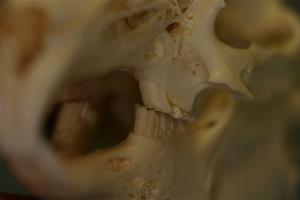
The incisors and cheek teeth of rabbits are called aradicular hypsodont (high-crowned
teeth and enamel which extends past the gum line) teeth. This is sometimes referred to as an elodent dentition.
These teeth grow or erupt continuously. The growth or eruption is held in balance
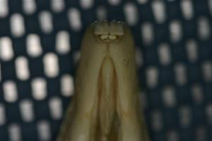
by dental abrasion from chewing a diet high in fiber. Incisors Familiarity with the incisor anatomy is important if the
veterinarian is considering extraction of these teeth. The upper incisors are
paired teeth on the left and right sides.
The front incisor is larger than the distal smaller (peg) tooth. The larger incisor is in the formation of a
half circle. The vestibular aspect of these incisors have a deep developmental
groove. The smaller peg incisor has a
broader curvature and is roughly half as long as the larger incisor. The mandibular incisor is longer than the
anterior upper incisor and its curvature is broader. The apical end of the mandibular incisor is
orientated lingual to the first mandibular cheek tooth (premolar).
Why would I take
the time to discuss the anatomy of the cheek teeth?
I believe an
understanding of this dental anatomy is critical to performing occlusal adjustments effectively. Understanding the normal anatomy allows the
veterinarian to recreate near normal anatomic dental arch relationships by
properly performing occlusal adjustment. The teeth of the rabbit are heterodont and diphydont. Heterodont teeth are simply teeth of different
types as opposed to teeth of the same type, called homodont. Rabbits have incisor teeth and cheek
teeth. The cheek teeth include both
premolars and molars. Rabbits do not
have canine teeth as in cats, dogs, ferrets and hedgehogs. Rabbits have a diphydont
dentition since they have deciduous (primary) and secondary (adult) teeth.
Anatomic Dental
Formula
2(i2/1 c0/0
m3/2)= 16 deciduous teeth 2(I2/1 C0/0 P3/2
M3/3)= 28 permanent teeth Cheek teeth
occlusion
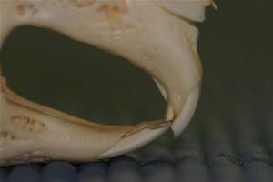
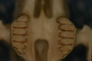
There is a
clear jaw width disparity when viewing the cheek teeth from an occlusal view. The
upper and lower jaw relationship is anisognathic. The mandibular dental arch consists of
premolars and molars orientated in a straight line. The upper dental arch (maxillary) consists
of premolars arranged in a lateral convex curved orientation. The dental arches have a slightly convex
curvature in the vertical plane. The
mandibular dental arch curves toward the buccal side and the maxillary arch
curves toward the tongue and the palate as viewed in the vertical plane. The jaws are anisognathic. The
mandibular dental arches are positioned slightly lingual to the maxillary
dental arches. The buccal edges of the
caudal mandibular molar cheek teeth, contact the palatal aspect of the
opposing maxillary molar cheek teeth. Dental attrition
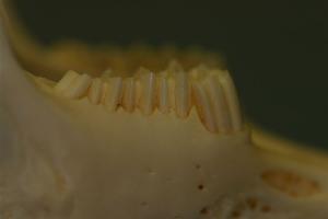
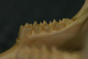
of rabbit cheek teeth
Dental
attrition is critical to the oral health of rabbits since their teeth
continuously grow. The buccal
surfaces of the mandibular cheek teeth wear more quickly than the lingual
aspects. The palatal-lingual aspect of
the maxillary cheek teeth wear more than the buccal aspects of these teeth. The anisognathic relationships of the upper and lower jaws causes the occlusal wear patterns and the development of the occlusal plane. The
veterinarian must be aware that the maxillary cheek teeth are normally longer
at the buccal aspect and shorter at the palatal-lingual aspect. The mandibular cheek teeth are normally
shorter at the buccal aspect and longer at the lingual aspect. The maxillary and mandibular cheek teeth
meet at approximately 15 degrees from horizontal (or level bite) to form the occlusal plane.
The purpose of occlusal adjustment is to
recreate this occlusal plane as accurately as
possible. There is less attrition
(wear) at the rostral and caudal aspects of the
upper dental arches and more in the middle.
The mandibular dental arches adapt to the maxillary dental attrition. Cheek teeth shape
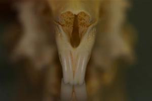
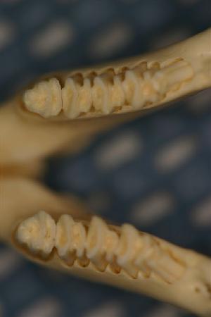
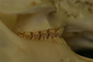
Occlusal aspect
The occlusal cusp surfaces of the cheek teeth
have enamel folds or ridges formed by enamel.
The troughs consist of dentin and cementum. The shape of the occlusal aspect of rabbit
mandibular cheek teeth are very close to square. The buccal to lingual measurement is close
to the mesial to distal measurement. The maxillary shape of the cheek teeth near
the occlusal surface is somewhat rectangular. The buccal to palatal-lingual measurement
is larger than the mesial to distal measurement. lingual-palatal
and buccal appearance
The side views
of the mandibular cheek teeth have folds called embrasures. The buccal aspect of the
mandibular cheek teeth have deep logitudinal
embrasures and the lingual aspect has more shallow logitudinal
embrasures. These embrasures are
located near the distal third of the mandibular cheek teeth. The maxillary cheek teeth also have logitudinal embrasures; however, they run down the middle
of the upper cheek teeth and are somewhat shallower than in the mandibular
cheek teeth. In the upper dental arch,
the first premolar tooth and the last molar tooth do not have
embrasures. In the mandible the last
molar lacks the embrasure but the first premolar has two embrasures on the
buccal side and one on the lingual side. Apical aspect of
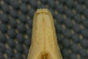
cheek teeth
The lower
cheek teeth apices diverge and protrude toward the ventral-lingual aspect of
the mandible. The upper cheek teeth
converge and project toward the buccal aspect of the maxilla.
|

e-mail: info@medirabbit.com
