![]()
Groei van het
oogbindvlies over het hoornvlies
pseudopterygium
Esther van Praag, Ph.D.
|
MediRabbit.com wordt uitsluitend
gefinancierd door gevers.
Elke donatie,
ongeacht hoe groot, wordt gewaardeerd en zal helpen bij de voortzetting van
het onderzoek van de medische zorg en de gezondheid van konijnen.
Bedankt
|

Waarschuwing: deze tekst bevat
foto’s die voor sommigen storend kunnen
zijn.
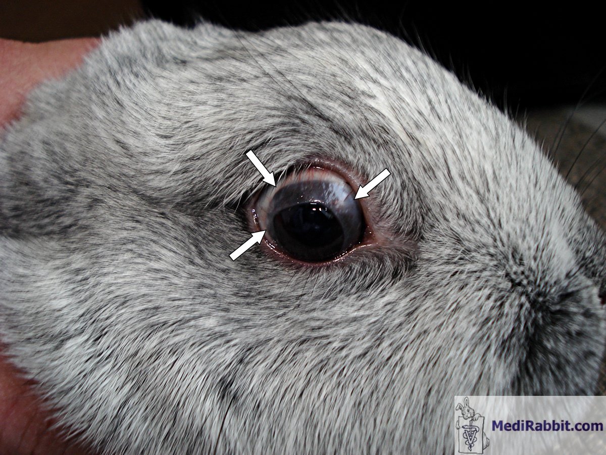
Er bestaan verschillende namen voor de afwijkende groei van het
oogbindvlies over het oogwit (sclera) naar en over het hoornvlies. Ras,
leeftijd of geslacht van de konijnen schijnen hierbij een rol te spelen sinds
meer mannelijke dwergkonijntjes, die tussen 5 en 12 manden oud zijn, door dit
probleem betroffen worden. Een mogelijke oorzaak kan ook de invloed van
ultraviolette bestraling zijn.
The etiology of the disease is unknown. It appears the result of an
inflammatory process that leads to the adherence of a fold of the conjunctiva
to the cornea, near the border of the cornea and the sclera (corneal limbus)
or more centrally. Breed, age, and sex of the rabbits seem to play a role, as
male dwarfs, aged between 5 and 12 months are more particularly affected. It
appears congenital in some cases. A further possible cause for pseudopterygium may be ultraviolet radiation.
Klinische tekenen
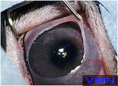
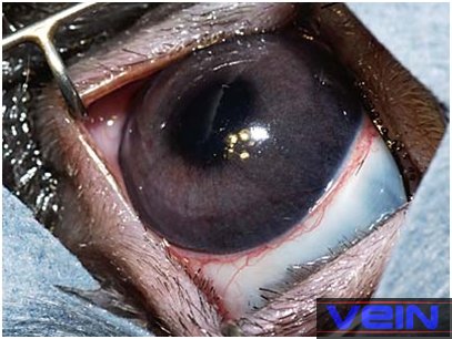
Pseudopterygium heeft specifiek het gedeelte “pseudo” in de naam,
doordat het overgroeiende membraan zich niet aan het onderliggende hoornvlies
hecht maar los erbovenop ligt. Als het verwijderd wordt, valt het meestal in
zijn normale positie terug. Het kan een klein gedeelte van het oog bedekken,
en zit er dan uit als een perifeere opaque ring, maar het kan ook het heel
oog bedekken, hetgeen tot blindheid leidt. When the membrane
is sectioned from the outer edge to the corneal limbus, it usually retracts
back to its normal position. The condition can be unilateral or affect both
eyes.
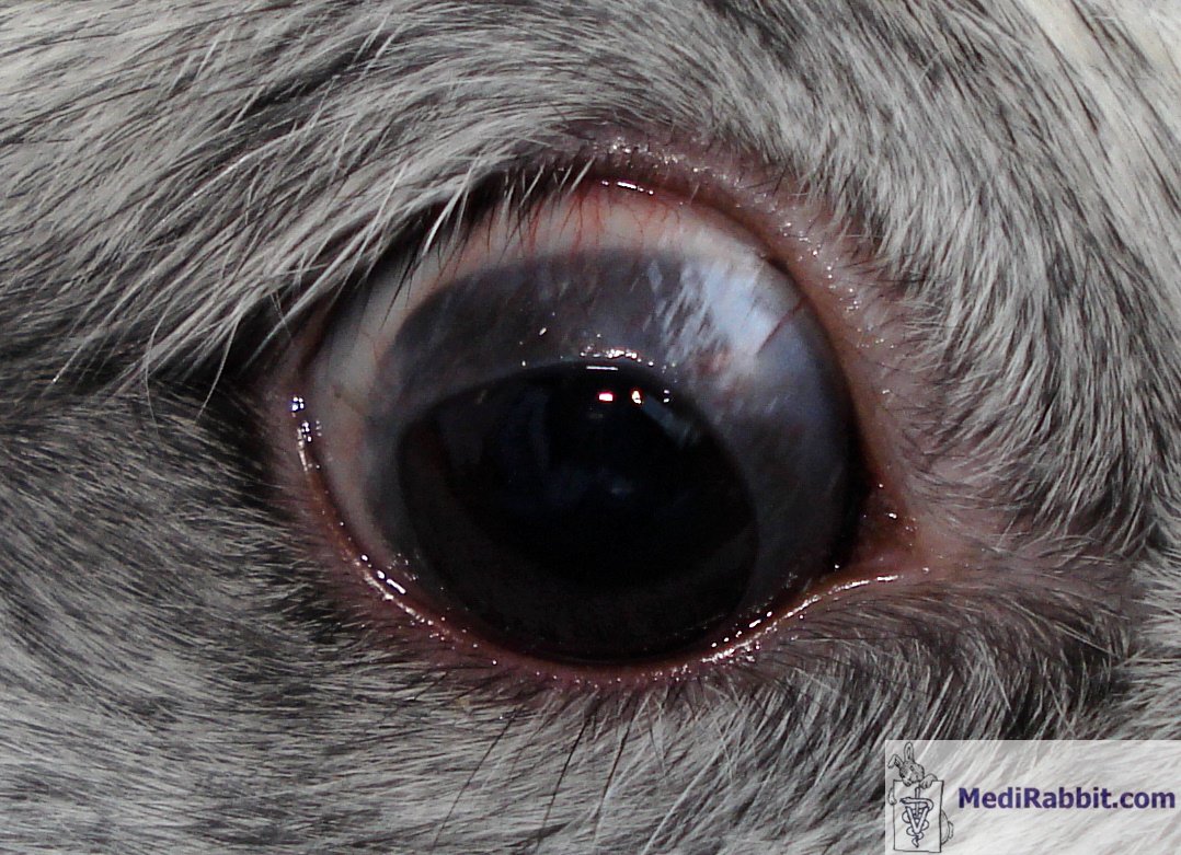
Meestal is pseudopetrygium pijnlijk if the
cornea is damaged by, e.g., a foreign body or a piece of hay stuck under the
membrane.
Stefan Röthlisberger
Het oogbindvlies membraan kan een klein gedeelte van het oog bedekkenArnbjer heeft een artikel over twee gevallen bij konijnen
gepubliceerd. In het eerste geval, is de oogbindvlies membraan met een scherp
instrument van de onderliggende hoorvlies verwijderd, gevolgd door injectie
van methylprednisolone acetaat onder in het bindvlies membraan. Het oog wordt
verder met steroïde/antibiotische oogdruppels gedurende 3 weken behandeld. In
het tweede geval was de nabehandeling met alleen maar antibiotische
oogdruppels, na de verwijdering van het membraan. Binnen weken was het weer
terug gegroeid.
Behandeling
Chirurgische verwijdering van het overgroeiende oogbindvlies leidt
vaak tot een terugkomende situatie. The condition can
be left untreated if it does not hinder the sight of the rabbit, and does not
cause pain.
Een betere behandeling schijnt het hechten van de membraam aan het ooglid to the sclera, or to the loose arching folds
connecting the conjunctival membrane lining the inside of the eyelid with the
conjunctival membrane covering the eyeball (fornix conjunctivae) on a
fully anesthesized rabbit. Dit kan met b.v.
oplossende draad zoals Dexon of Vicryl 5.0 of 6.0 gedaan worden. Het is
gevolgd door een plaatselijke behandeling met cyclosporine 0,2% en
corticosteroïden (b.v. dexamethasone 0.1%) gedurende een par weken.
Rarely, the condition may become chronic, with repeated re-growth of
the membrane; life-long follow-up is needed to minimize regrowth of the conjunctival
membrane.
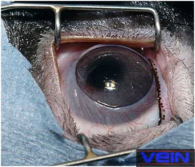
Akira Yamanouchi
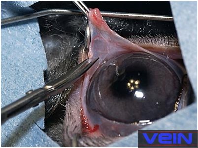
Pre-surgical
preparation for the removal of the membrane growing over the cornea
Akira Yamanouchi
The membrane is removed, by cutting it in 4 to 6
segments, from the edge back to the conjunctival bulbar tissue. Once placed
in it normal position, the membrane is sutured to
the eyelid, the sclera or the fornix conjunctivae.
Sutures can be removed after 3 weeks, or left in
place.
Bedanktuiging
Een woord van dank gaat naar Akira
Yamanouchi voor de toestemming de foto’s uit VEIN (Veterinary Exotic
Information Network, https://vein.ne.jp/). betreffende konijnen ziektes te mogen
gebruiken.
Ook een speciale dank
aan Louise en Arie
van Praag (Zwitserland), voor hun hulp bij de editie van
teksten in het Nederlands.
Verdere informatie
Arnbjer, J. Pseudopterygium in a pygmy rabbit. Vet. Med. Small Anim. Clin. 74,737-738 (1979).
Bourne D.
Aberrant conjunctival overgrowth in rabbits.
https://wildlife1.wildlifeinformation.org/S/00dis/Miscellaneous/AbConjunctOvergrowthRabbit.html
Delaney, K.H.
Diagnostic exercise: Apparent corneal occlusion in a New Zealand white
rabbit. Contemp. Top. Lab. Anim. Sci. 34,76-77
(1995).
DuPont, C.,
Carrier, M. & Gauvin, J. Bilateral precorneal
membranous occlusion in a dwarf rabbit. J. Small Exotic
Anim. Med. 3,41-44 (1995).
Fehr, M. Eye
anomalies in dwarf rabbits. [German].
Kleintierpraxis 29, 129-130, 132 (1984).
Matros, L.E., Ansari,
M.M. & Van Pelt, C.S. Eye anomaly in a dwarf rabbit. Avian Exotic Pract. 3,13-14 (1986).
Roze, M., Ridings, B.
& Lagadic, M. Comparative morphology of epicorneal conjunctival membranes in rabbits and human
pterygium. Vet. Ophthalmol. 4,171-174 (2001).
Schoofs, S. &
Hanssen, P. Epicorneal conjunctival membrane in the
rabbit: a clinical case and surgical therapy. Vlaams Diergeneeskundig
Tijdschrift 67,344-346 (1998).
Wagner, F., Heider, H.J., Gorig, C. &
Fehr, M. Ophthalmic diseases in dwarf rabbits. Part 1: eye examination,
anatomy, diseases of the eyelids, the conjunctiva and of the nasolacrimal
duct. [German]. Tierarztl. Prax. 26,205-210 (1998).
|
e-mail: info@medirabbit.com