![]()
Female reproductive tract and ovariohysterectomy
(spay surgery)
Esther van Praag, Ph.D.
|
MediRabbit.com is funded solely by the
generosity of donors.
Every donation, no matter what
the size, is appreciated and will aid in the continuing research of medical care
and health of rabbits.
Thank you
|

WARNING: this file contains
pictures of the surgical procedure that may be distressing for people.
|
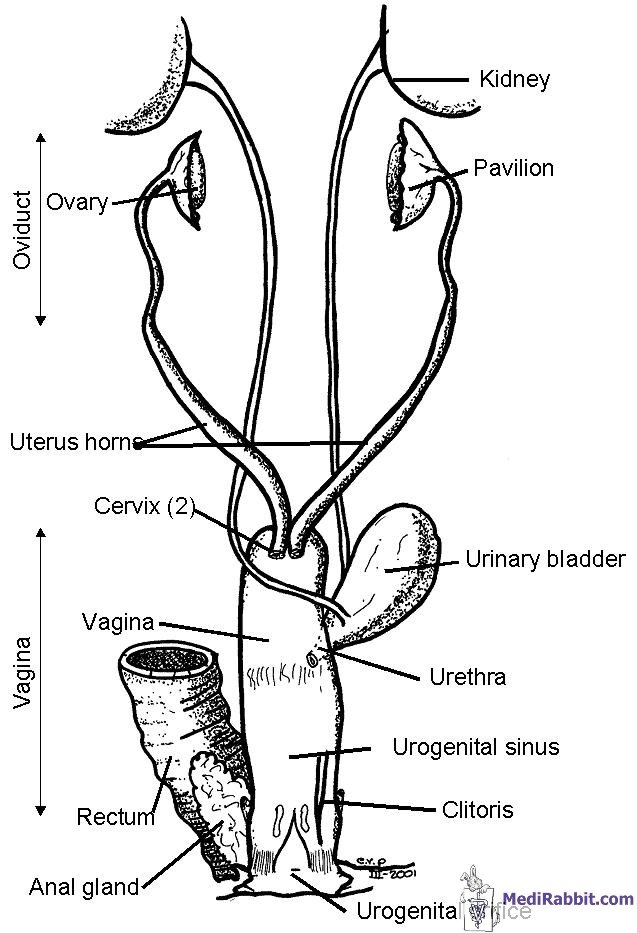
The
reproductive organ of the female rabbit is considered primitive. Indeed, the
split two-horned system is only encountered in monotreme
egg laying mammals and the lagomorphs (pika, hare
and rabbit). The organ is held in place by a broad ligament that is anchored
at 4 points under the vertebral column. Sex
differentiation occurs during the embryonic phase, on the 16th day
post-fertilization. The ovaries grow from an aggregate of cells that is lying
near the original testes. The development of the ovaries is accompanied by
the degeneration of the testes. The
development of the ovules (female reproductive cell) starts around the 21st
day and continues till birth, around the 30th day. The first ova and
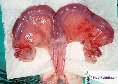
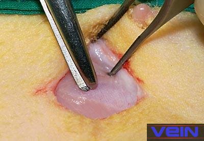
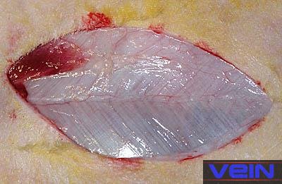
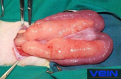
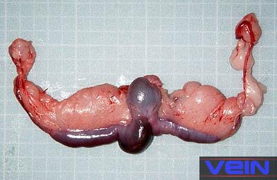
follicles start to develop only 13 days after birth. The reproductive organ of the female
rabbit is duplex: the uterus is formed by two independent horns, split over
their whole length (± 7 cm). Each horn possesses its own cervix. The ovaries,
ellipsoid bodies that have a maximal length of 1-1.5 cm, are located at the
end of the uterus, right under the kidneys. They are hidden by the mesometrium (portion of the broad ligament that separates
and encloses the uterus) and fat.
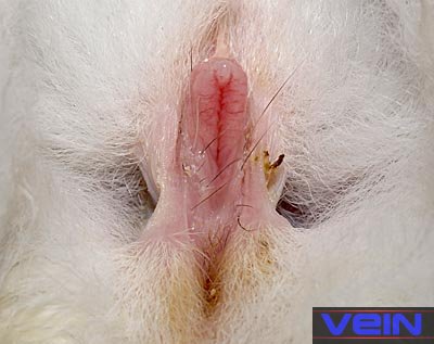
The
vagina does not present any particularities. This part of the reproductive
tract is large, with the urethra joining halfway, at the level of the vaginal
vestibule. At the end of the vagina, the glands of Bartholin and preputial
glands can be recognized. The
age at which sexual maturity is reached depends on the size and the breed:
while small and middle sized rabbits become adult between 4 and 6 months, it
may take between 5 to 8 months for giant breeds. As a rule, it is considered
that a rabbit is adult and able to reproduce when it has reached 75 to 80% of
its adult size. Female
rabbits do not have an estrus (heat) cycle, as do other small animals like dogs
or cats, but are called induced ovulators.
Ovulation is spontaneous; it happens 9 to 13 h. after copulation. A certain cycle does nevertheless
exist. The presence of the estrogen hormone will influence the size and the
color of the vulva. Most female rabbits are more prone to mate when their
vulva is colored reddish/purple. It is no clear indication though, as some
female rabbits, called “reflex ovulators”, will
mate when their vulva is pale and small.
Spay surgery
The
spay of female rabbits is recommended in order to avoid unwanted litters,
pseudo-pregnancies and reduce difficult behavior, like the marking of its
territory with strong smelling urine, aggressive attitude and mounting of
objects or human body parts like feet and arms. Further medical reasons to
favor of a spay surgery include: •
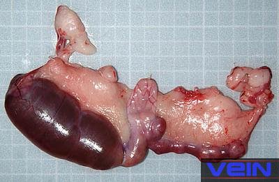
Difficult to treat bacterial infection or pyometra; • Neoplasia of the ovaries or uterus
(e.g. adenocarcinoma), a problem frequently seen in unspayed
older rabbits; •
Abnormal swelling of the wall of the uterine artery
(uterine aneurysm). Ovariohysterectomy
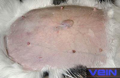
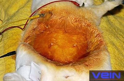
is a surgical procedure done under general anesthesia, in a sterile
environment, with sterile surgical instruments and a preparation of the skin.
Indeed, any post-surgical infection becomes a medical emergency. A spay is usually done around the age of
5 months, or when the rabbit is sexually mature. At a younger age, the
surgery is complicated by the difficulty to locate the very thin uterus and
the very small ovaries. A spay at immature age may also have a dramatic
effect on the proper calcium absorption by the bones.
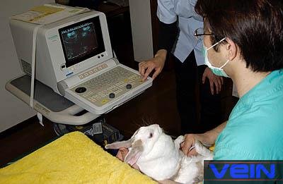
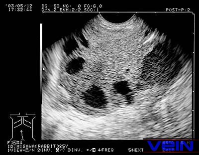
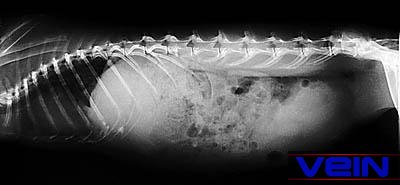
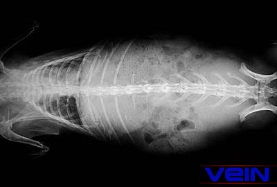
When a spay surgery is done in an
older rabbit, it is advisable to do an ultrasound prior to surgery in order
to rule out the presence of tumors, and, if found, to check for the presence
of metastasis (e.g. in the lungs) before the surgery.
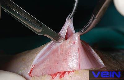
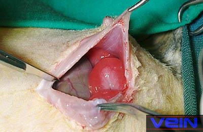
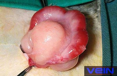
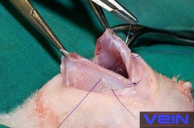
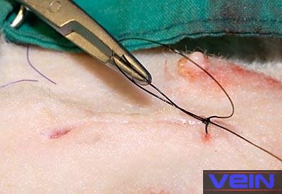
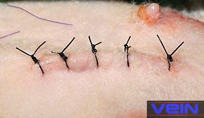
Various surgical approaches are
available for ovariohysterectomy in rabbits, described in books, videos, or
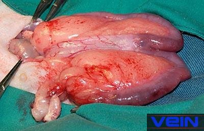
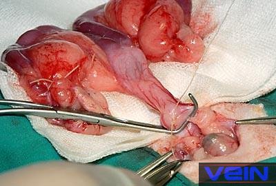
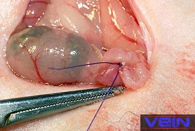
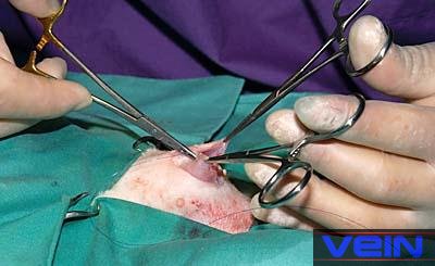
scientific articles. Video and step by step
illustration of a spay surgery in a female rabbit
Post
surgical care
The administration of pain medication for several days after the
surgery is highly recommended. Pain indeed induces hormonal and physiological
responses, which slow down the activity of the digestive tract, delay food
intake and recovery. The presence of blood in the urine during 24 to 48 hrs. post-surgery is sometimes observed. Rabbits have a propensity to form intra-abdominal
adhesion post-operatively. These complications can be reduced by the
administration of verapamil (200 μg/kg,
SC, tid for 3 days). Studies showed that the
NSAID pain drug ibuprofen has the same preventive effect against adhesion as
verapamil. The rabbit should furthermore be confined for 7 to
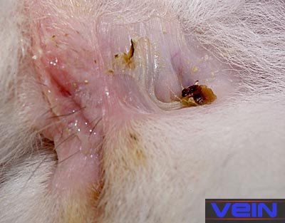
10 days, and kept separated from other rabbits up to 10 days post-surgery. Healthy and diseased
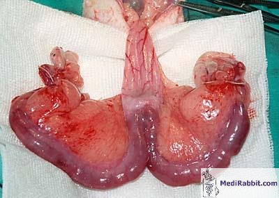
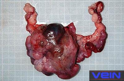
female reproductive organs
Acknowledgement
Many thanks to Akira Yamanouchi, for
the permission to use pictures from VEIN (Veterinary
Exotic Information Network). Many thanks also to Debbie Hanson
(USA) for the spay videos. a Further Information
Atherton
J, Griffiths L, Williams A. Cystic mastitis disease in the female rabbit. Vet
Rec. 1999 Nov 27;145(22):648. Bray MV,
Weir EC, Brownstein DG, Delano ML. Endometrial venous aneurysms in three New
Zealand white rabbits. Lab Anim Sci. 1992 Aug;42(4):360-2. Cao T,
Shirota T, Ohno K, Michi
KI. Mineralized bone loss in partially edentulous trabeculae of ovariectomized rabbit mandibles. J Periodontal Res. 2004
Feb;39(1):37-41.
Gilsanz V, Roe TF, Gibbens
DT, Schulz EE, Carlson ME, Gonzalez O, Boechat MI.
Effect of sex steroids on peak bone density of growing rabbits. Am J Physiol.
1988 Oct;255(4 Pt 1):E416-21. Hussein
SA, Azab ME, Abdel-Maksoud
H. Metabolic changes concerning the effect of castration on some blood
constituents in male rabbits. Dtsch Tierarztl Wochenschr. 1999 Mar;106(3):113-8. Jenkins
JR. Surgical sterilization in small mammals. Spay and castration. Veterinary
Clin North Am Exot Anim Pract. 2000 Sep;3(3):617-27, v. Review. Lester-Cockx L. Neutering pet rabbits. Vet Rec. 1999 Mar 6;144(10):271. Meredith
A, Redrobe S, Keeble E. Neutering pet rabbits. Vet
Rec. 1999 Mar 20;144(12):328. Millis DL,
Walshaw R. Elective castrationsd
and ovariohysterectomies in pet rabbits. J. Am. Anim
Hosp. Assoc. 1992: 491-497 Quesenberry KE, Carepenter
JW, Quesenberry P. Ferrets, Rabbits and Rodents:
Clinical Medicine and Surgery Includes Sugar Gliders and Hedgehogs, Elsevier
Health, 2004. Raftery A. Uterine adenocarcinoma in
pet rabbits. Vet Rec. 1998 Jun 20;142(25):704. Smith K. Rabbit
Health in the 21st Century. A Guide for Bunny Parents, Second Edition https://rabbithealth101.com/ Harcourt-Brown
F. Textbook of Rabbit Medicine, UK: Butterworth-Heinemann, 2001. Flecknell P ,
editor. BSAVA Manual of Rabbit Medicine and Surgery, Gloucester, UK: British
Small Animal Veterinary Association, 2000. Sommerville LM.
Treatment of a uterine adenocarcinoma in a domestic rabbit by
ovariohysterectomy. Vet Rec. 1998 May 16;142(20):550-1. |
||||||||||||||||||||||||||||||||||

e-mail: info@medirabbit.com
