![]()
Endometritis, Orchitis and Pyometra
Esther van Praag, Ph.D.
|
MediRabbit.com is funded solely by the
generosity of donors.
Every donation, no matter what
the size, is appreciated and will aid in the continuing research of medical care
and health of rabbits.
Thank you
|

Warning: this file
contains pictures that may be distressing for people.
|
Endometritis = inflammation
of the endometrium, the mucous
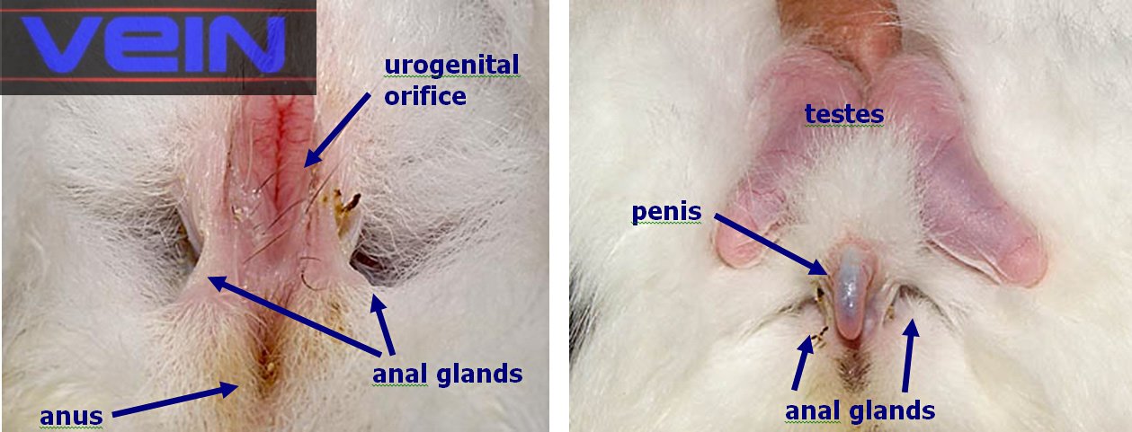
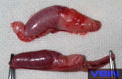
membrane lining the uterus. Orchitis = inflammation of the testis. Pyometra = literally meaning “pus in the uterus”,
or purulent inflammation of the uterus. Both
female and male rabbits can be affected by infections of the urogenital
tract. The causing agents are generally Pasteurella multocida, and Staphylococcus
aureus, though other bacteria should not be ruled out. The development of
the disease depends on the general resistance of the host, and the virulence
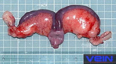
of the bacteria. Endometritis, orchitis,
and pyometra are disorders commonly found in
rabbits. Generally, the affections are
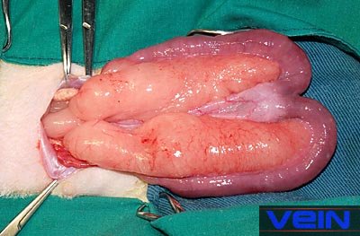
chronic; this is due to the dough-like nature of the exudates, which the
uterus is unable to drain properly. This leads to a massive enlargement of
the uterus, and the risk of rupture.
Pyometra often relates to the presence of Pasteurella
multocida. It leads to the formation of abscesses in the ovaries. The
uterus is dilated, and generally filled with pus. Acute infections are often
accompanied by a vaginal discharge. Both breeding and virgin does can be
affected. Three possible ways of transmission are possible: • retrograde
transmission from the nasal cavity to the genital area during coprophagy, by a female rabbit infected by P.
multocida, • transmission
during the kindling (birth) procedure, • venereal transmission, when an infected female mates with
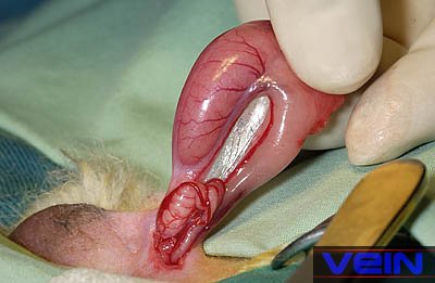
an uninfected male rabbit, or vice-versa. In males, the testicles and epididymis
are often swollen during the acute phase of the infection; in a later stage,
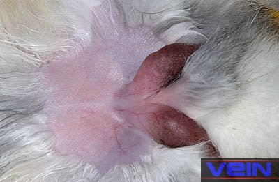
external or internal abscesses appear in the testicle. In rare cases, the
infection limits itself to the membrane covering the penis, a condition
called balanoposthitis. The membrane is inflamed,
and covered with pus. The infection can be transmitted to female rabbits,
during attempts to mount or copulate Clinical signsThe
rabbit is often depressive, lethargic, anorexic, and shows general weakness. Typically,
the abdomen is enlarged, the uterus is distended, and vaginal discharge may
be observed. Sometimes it is accompanied by polydipsia (excessive intake of
water), and/or polyuria (excessive excretion of urine). Male
rabbits generally are feverish, show irregular appetite, lose weight, and are
infertile. DiagnosisIn the female rabbit, an enlarged uterus should be
examined carefully, as the distended membrane is fragile. Palpation of the
uterus helps determine its consistency: smooth or doughy. If an enlarged abdomen is diagnosed, X-ray
radiographs of the lower abdomen may provide additional information.
Ultrasound imaging help to rule out other uterine
disorders like polyps, masses, and cystic changes. Analysis of serum biochemistry; amyloid deposition
in the kidney is often a consequence of chronic infection of the uterus.
Blood analysis can reveal mild normocytic anemia, hypercholesterolemia, heterophilia (the presence of granular leukocytes), and monocytosis (presence of mononuclear phagocytic leukocyte).
See: Blood biochemistry of rabbits Cervical/mucus drainage can be used for a Gram stain. When in the
presence of open endometritis, a guarded deep vaginal/cervical swab can be
taken in order to determine the causing agent of the infection. Pasteurella
multocida, and Staphylococcus aureus, more rarely Listeria sp, are often involved. Confirmation of the diagnosis can be done through laparotomy (incision
through the abdominal wall) or ovariohysterectomy.
In females, the diagnosis must differentiate from
other causes such as uterine adenocarcinoma, hydrometra, endometrial venous
aneurysm (sac formed by abnormal dilatation of the vein wall), and dystocia
(abnormal labor). In males, a differential culture should be made to differentiate
between a bacterial infection, a viral infection (e.g. myxoma
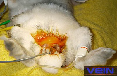
virus), spirochetosis or rabbit syphilis TreatmentFor female rabbits, ovariohysterectomy is
recommended. It is, however, important to stabilize the animals before the operation;
treatment includes the administration of antibiotics, and fluid therapy.
During the removal of the uterus, samples should be collected for bacterial
culture, and antibiotic sensitivity should be determined. After the
operation, IV fluid therapy, broad-spectrum antibiotics, and pain medication
must be administrated. In males, the treatment of choice is castration, and
post-surgical administration of pain medication, and antibiotics. Finally, it is important to check the animals for abscesses located
elsewhere.
AcknowledgementThanks are due to Akira Yamanouchi, for the
permission to use pictures from VEIN (Veterinary Exotic Information Network, https://vein.ne.jp/). Further InformationBjotvedt G, Bertke EM, Hendricks GM. Peritonitis due to Pasteurella
multocida in a rabbit. Vet Med Small Anim Clin. 1979; 74(2):215-6.
Flatt RE. Pyometra
and uterine adenocarcinoma in a rabbit. Lab Anim
Care. 1969; 19(3):398-401 Flecknell PA BSAVA
Manual of Rabbit Medicine and Surgery, British Small Animal Veterinary; 2000. Fountain S, Holland MK, Hinds LA, Janssens PA, Kerr PJ. Interstitial orchitis
with impaired steroidogenesis and spermatogenesis in the testes of rabbits
infected with an attenuated strain of myxoma virus.
J Reprod Fertil. 1997;
110(1):161-9. Hobbs BA, Parker RF. Uterine
torsion associated with either hydrometra or endometritis in two rabbits. Lab
Anim Sci. 1990; 40(5):535-6. Hofmann JR Jr,
Hixson CJ. Amyloid A protein deposits in a rabbit with pyometra.
J Am Vet Med Assoc. 1986; 189(9):1155-6. Hoffmann JA. Orchitis
and epididymitis of traumatic origin in a male rabbit. Dtsch
Tierarztl Wochenschr.
1963; 70(18):524. Johnson JH, Wolf AM. Ovarian abscesses and pyometra
in a domestic rabbit. J Am Vet Med Assoc. 1993; 203(5):667-9. Peters M, Scheele G. Listeriosis in a rabbitry. Dtsch
Tierarztl Wochenschr. 1996; 103(11):460-2. German. Schmidt S, Schrag D, Giese B.
Ultrasonic diagnosis in gynecology in small animals. Tierarztl
Prax. 1986; 14(1):123-41. Tumboh-Oeri AG, Roberts
TK. Immunological and morphological consequences of vasectomy in the rabbit. Experientia. 1979; 35(5):675-6. |
e-mail: info@medirabbit.com
