![]()
Myxomatosis in rabbits
Esther van
Praag, Ph.D.
|
|
MediRabbit.com is
funded solely by the generosity of donors. Every
donation, no matter what the size, is appreciated and will aid in the
continuing research of medical care and health of rabbits. Thank you |

Warning: this file contains pictures that may be distressing to some persons
Sanarelli
first recognized the myxomatosis disease in 1896, in Uruguay, where it causes
sporadic lethal infections in the American cottontail species (Sylvilagus
sp.). The virus has spread over the entire American continent, and has become
endemic in some regions (Chili, in O. cuniculi; Western USA, in Sylvilagus
bachmani).
It was soon discovered that the
European rabbit, (Oryctolagus cuniculi) was very sensitive to this
virus, causing severe skin abscesses, infections, and ultimately death. In the
1950se, the myxomatosis virus was introduced and spread among wild rabbits in
Australia, in order to reduce its population. This operation decimated almost
all the wild rabbit population, except a few individuals that seemed
resistant to this virus. The surviving rabbits started to reproduce offspring
and colonized the country anew. In Europe, the virus spread rapidly and has
now become endemic in some regions, which are populated by the European
rabbit.
The
lagomorph’s groups largely affected by the myxoma virus are the European
rabbit (Oryctolagus cuniculi), the European hare (Lepus europaeus),
the Brush cottontail (S. bachmani) and the eastern cottontail (S.
floridanus).
Myxomatosis
is caused by a virus belonging to the family of the Poxviridae, and is a type
species of the genus Leporipoxvirus. The later comprises close related
viruses that affect he American cottontail rabbit (Sylvilagus sp.) and
the “hare fibroma virus”, among others. All these viruses lead to the
development of tumors of the skin connective tissues (fibroma). Various
strains exist. Some are very virulent (e.g. Standard laboratory, Lausanne,
California), others manifest their presence chronically. Genetic
studies show a relationship between the myxoma and the Shope fibroma virus Blood sucking insects (fleas, mosquitoes, lice and
mite) are efficient mechanical vectors of this disease. It was observed that
the virus is present in the mouth’s parts of the rabbit flea Spilopsyllus
cuniculi, where it can survive over 100 days, independently of the
environmental conditions. It is furthermore speculated that the disease may
spread from one rabbit to another during skin and fur contact.
Clinical signs
The
development of myxomatosis follows the typical pattern of a poxvirus infection.
Once inoculated in the skin, the virus starts to reproduce in the skin and
local lymph nodes. The virus is then spread through the body. The viruses are
then spread throughout the body (viremia) and into the skin.
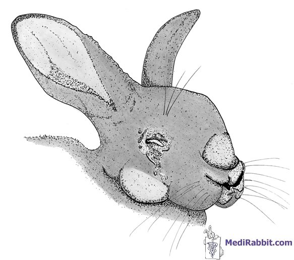
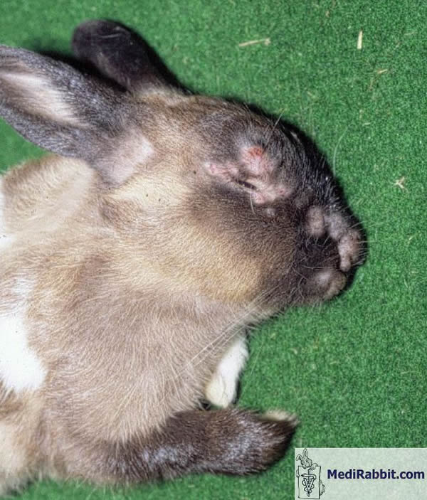
The
first evident signs of the disease appear 3 days after the infection:
swelling (edema) of the eyelids, followed by the lips, genital organs and
purulent conjunctivitis. At later stages of the disease, the rabbit becomes
blind. The disease is usually fatal between day 8 to
15 after the infection with the virus.
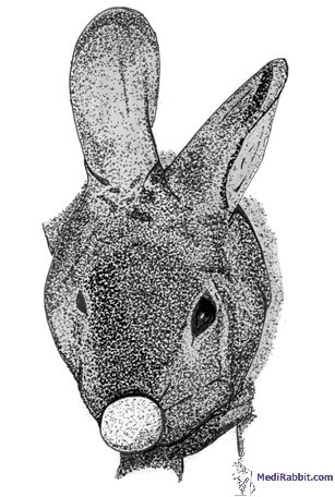
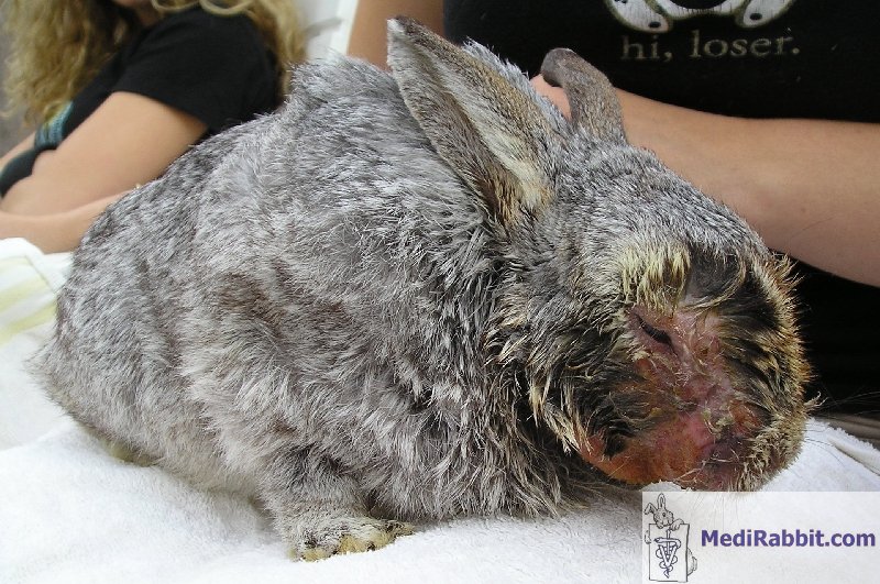
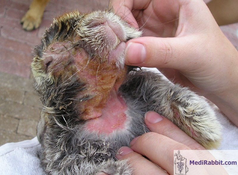
In
the chronic form of the disease, the most prominent signs are the formation
of skin tumors, called myxoma, on the ears, nose and limbs. These tumors will
resorb by themselves after some time.
A
side effect of the chronic form of myxomatosis is the development of
secondary bacterial infection. Pneumonia caused by Pasteurella sp. or Staphylococcus
aureus is often observed. It is accompanied by respiratory distress
(dyspnea).
Diagnosis
Although
the disease depends on the strain of myxoma virus, it is usually severe and
almost always fatal. The
clinical symptoms are sufficient for diagnosis. One must keep in mind though,
that early stages of the spirochetosis disease (caused by the parasite Treponema
sp., affecting the perianal parts of the rabbit) look similar to those of
myxomatosis. Indeed, tumors of those diseases show close similarities, so
spirochetosis and myxomatosis must be carefully differentiated from each
other. Myxomatosis
should furthermore be differentiated from an upper respiratory infection,
like e.g. Pasteurellosis. In the later, no swelling is observed in the
perianal region, on the contrary to myxomatosis. In the
case myxomatosis is chronic, it is recommended to do a biopsy and check it
for the presence of viruses. Treatment
If a rabbit is affected by the aggressive form of myxomatosis, its
chances of survival are close to zero. It is then recommended to humanely put
the affected animal to sleep. If treatment is chosen, intensive care over a longer period of time is
needed. It is important to keep the sick rabbit in a warm environment
(21-22°C). Eyes and ears must be regularly cleaned. As much fluids and food
should be given to the rabbit as possible, even if the rabbit is drinking
good amounts of water by itself. Skin tumors can be removed surgically. Unfortunately, secondary complications often appear. The most common
one are respiratory disease and pneumonia, due to secondary infection by Pasteurella
sp. or Staphylococcus sp. Rabbit that suffer a chronic form of myxomatosis recover by
themselves. Antibiotics can be given to avoid respiratory complications. In regions where myxomatosis is endemic and present among the wild
rabbit or cottontail population, prevention of the disease in pet rabbits is
is possible by regular vaccination. This is not available in all countries.
Depending
on the vaccine used and the age or breed of rabbits, vaccinated rabbits may
develop a mild to serious form of the disease. In rare cases, the rabbit must
be put to sleep.
For detailed information on myxomatosis in rabbits,
by E. van Praag, A. Maurer and T.
Saarony, 408
pages, 2010.
Acknowledgement
A special thanks to Denise Baart, for
sharing the pictures of her rabbit Bucks. Further Reading
Best SM, Collins SV, Kerr PJ.
Coevolution of host and virus: cellular localization of virus in myxoma virus
infection of resistant and susceptible European rabbits. Virology. 2000;
277(1):76-91. Boag B. Observations on the
seasonal incidence of myxomatosis and its interactions with helminth parasites
in the European rabbit (Oryctolagus cuniculus). J Wildl Dis. 1988;
24(3):450-5. Boag B, Lello J, Fenton A,
Tompkins DM, Hudson PJ. Patterns of parasite aggregation in the wild European
rabbit (Oryctolagus cuniculus). Int J Parasitol. 2001; 31(13):1421-8. Calvete C, Estrada R, Villafuerte
R, Osacar JJ, Lucientes J. Epidemiology of viral haemorrhagic disease and
myxomatosis in a free-living population of wild rabbits. Vet Rec. 2002;
150(25):776-82. Chapple PJ, Lewis ND. Myxomatosis
and the rabbit flea. Nature. 1965; 207(995):388-9. Chapuis JL, Chantal J, Bijlenga G.
Myxomatosis in the sub-antarctic islands of Kerguelen, without vectors,
thirty years after its introduction. C R Acad Sci III. 1994; 317(2):174-82. Duclos P, Caillet J, Javelot P. Aerobic
bacterial flora of the nasal cavity of rabbits. Ann Rech Vet.
1986; 17(2):185-90. Edmonds JW, Nolan IF, Shepherd RC,
Gocs A. Myxomatosis: the virulence of field strains
of myxoma virus in a population of wild rabbits (Oryctolagus cuniculus
L.) with high resistance to myxomatosis. J Hyg (Lond). 1975; 74(3):417-8. Flowerdew JR, Trout RC, Ross J.
Myxomatosis: population dynamics of rabbits (Oryctolagus cuniculus
Linnaeus, 1758) and ecological effects in the United Kingdom. Rev Sci
Tech. 1992; 11(4):1109-13. Fountain S, Holland MK, Hinds LA,
Janssens PA, Kerr PJ. Interstitial orchitis with impaired steroidogenesis and
spermatogenesis in the testes of rabbits infected with an attenuated strain
of myxoma virus. J Reprod Fertil. 1997; 110(1):161-9. Ghram A, Benzarti M, Amira A,
Amara A. Myxomatosis in Tunisia: seroepidemiological study in the Monastir
region (Tunisia). Arch Inst Pasteur Tunis. 1996; 73(3-4):167-72. Gorski J, Mizak B, Chrobocinska M.
Control of rabbit myxomatosis in Poland. Rev Sci Tech. 1994; 13(3):869-79. Jiran E, Sladka M, Kunstyr I.
Myxomatosis of rabbits--study of virus modification. Zentralbl Veterinarmed
B. 1970; 17(3):418-28. Joubert L, Tuaillon P, Larbaigt G.
Serologic and allergologic relationship between rabbit myxomatosis and
fibromatosis viruses. Conglutination reaction and homologous and heterologous
hypersensitivity. Bull Acad Vet Fr. 1970; 43(6):259-76. Joubert L, Oudar J, Mouchet J,
Hannoun C. Transmission of myxomatosis by mosquitoes in Camargue. Preeminent
role of Aedes caspius and Anopheles of the maculipennis group.
Bull Acad Vet Fr. 1967; 40(7):315-22. Kerr PJ, Merchant JC, Silvers L,
Hood GM, Robinson AJ. Monitoring the spread of myxoma virus in rabbit Oryctolagus
cuniculus populations on the southern tablelands of New South Wales,
Australia. II. Selection of a strain of virus for release. Epidemiol Infect.
2003; 130(1):123-33. Kerr PJ, Best SM. Myxoma virus in
rabbits. Rev Sci Tech. 1998; 17(1):256-68. Lawton MP. Myxomatosis vaccine.
Vet Rec. 1992; 130(18):407-8. Licon Luna RM. First report of
myxomatosis in Mexico. J Wildl Dis. 2000; 36(3):580-3. Marcato PS, Simoni P.
Ultrastructural researches on rabbit myxomatosis. Lymphnodal lesions. Vet
Pathol. 1977; 14(4):361-7. Marlier D, Mainil J, Linde A,
Vindevogel H. Infectious agents
associated with rabbit pneumonia: isolation of amyxomatous myxoma virus
strains. Vet J. 2000; 159(2):171-8. Merchant JC, Kerr PJ, Simms NG,
Robinson AJ. Monitoring the spread of myxoma virus in rabbit Oryctolagus
cuniculus populations on the southern tablelands of New South Wales,
Australia. I. Natural occurrence of myxomatosis. Epidemiol Infect. 2003;
130(1):113-21. Merchant JC, Kerr PJ, Simms NG,
Hood GM, Pech RP, Robinson AJ. Monitoring the spread of myxoma virus in
rabbit Oryctolagus cuniculus populations on the southern tablelands of
New South Wales, Australia. III. Release, persistence and rate of spread of
an identifiable strain of myxoma virus. Epidemiol Infect. 2003;
130(1):135-47. Nash P, Barrett J, Cao JX,
Hota-Mitchell S, Lalani AS, Everett H, Xu XM, Robichaud J, Hnatiuk S, Ainslie
C, Seet BT, McFadden G. Immunomodulation by viruses: the myxoma virus story.
Immunol Rev. 1999; 168:103-20. Omori M, Banfield WG. Shope
fibroma and rabbit myxoma factories: electron microscopic observations. J
Electron Microsc (Tokyo). 1970; 19(4):381-3. Patterson-Kane J. Study of
localised dermatosis in rabbits caused by myxomatosis. Vet Rec. 2003;
152(10):308. Patton NM, Holmes HT. Myxomatosis
in domestic rabbits in Oregon. J Am Vet Med Assoc. 1977; 171(6):560-2. Regnery DC. The epidemic potential
of Brazilian myxoma virus (Lausanne strain) for three species of North
American cottontails. Am J Epidemiol. 1971; 94(5):514-9. Regnery DC, Miller JH. A myxoma virus
epizootic in a brush rabbit population. J Wildl Dis. 1972; 8(4):327-31. Robinson AJ, Muller WJ, Braid AL,
Kerr PJ. The effect of buprenorphine on the course of disease in laboratory
rabbits infected with myxoma virus. Lab Anim. 1999; 33(3):252-7. Ross J. Myxomatosis and the
rabbit. Br Vet J. 1972; 128(4):172-6. Ross J, Tittensor AM. The
establishment and spread of myxomatosis and its effect on rabbit populations.
Philos Trans R Soc Lond B Biol Sci. 1986; 314(1167):599-606. Ross J, Sanders MF. The development
of genetic resistance to myxomatosis in wild rabbits in Britain. J Hyg
(Lond). 1984; 92(3):255-61. Ross J, Tittensor AM, Fox AP,
Sanders MF. Myxomatosis in farmland rabbit populations in England and Wales.
Epidemiol Infect. 1989; 103(2):333-57. Ross J, Sanders MF. Changes in the
virulence of myxoma virus strains in Britain. Epidemiol Infect. 1987;
98(1):113-7. Rothschild M. Myxomatosis and the
rabbit flea. Nature. 1965; 207(2):1162-3. Sellers RF. Possible windborne
spread of myxomatosis to England in 1953. Epidemiol Infect. 1987
Feb;98(1):119-25 Shepherd RC. Myxomatosis: the
occurrence of Spilopsyllus cuniculi (Dale) larvae on dead rabbit
kittens. J Hyg (Lond). 1978; 80(3):427-9. Shepherd RC, Edmonds JW.
Myxomatosis: the release and spread of the European rabbit flea Spilopsyllus
cuniculi (Dale) in the Central District of Victoria. J Hyg (Lond). 1979;
83(2):285-94. Sobey WR, Conolly D, Haycockp,
Edmonds JW. Myxomatosis. The effect of age upon survival of wild and domestic
rabbits (Oryctolagus cuniculus) with a degree of genetic resistance
and unselected domestic rabbits infected with myxoma virus. J Hyg (Lond).
1970; 68(1):137-49. Sobey WR, Conolly D. Myxomatosis:
passive immunity in the offspring of immune rabbits (Oryctolagus cuniculus)
infested with fleas (Spilopsyllus cuniculi Dale) and exposed to myxoma virus.
J Hyg (Lond). 1975; 74(1):43-55. Torres JM, Sanchez C, Ramirez MA,
Morales M, Barcena J, Ferrer J, Espuna
E, Pages-Mante A, Sanchez-Vizcaino JM. First field
trial of a transmissible recombinant vaccine against myxomatosis and rabbit
hemorrhagic disease. Vaccine. 2001; 19(31):4536-43. Trout RC, Ross J, Fox AP. Does
myxomatosis still regulate numbers of rabbits (Oryctolagus cuniculus
Linnaeus, 1758) in the United Kingdom? Rev Sci Tech. 1993; 12(1):35-8. Werffeli F. Observations on the
appearance of a clinically atypical manifestation of myxomatosis in wild and
domestic rabbits. Schweiz Arch Tierheilkd. 1967; 109(1):9-16. Williams RT, Dunsmore JD, Parer I.
Evidence for the existence of latent myxoma virus in rabbits (Oryctolaqus
cuniculus (L.)). Nature. 1972; 238(5359):99-101. Wunderwald C, Hoop RK, Not I,
Grest P. Myxomatosis in the rabbit. Schweiz Arch Tierheilkd. 2001;
143(11):555-8 Zuniga MC.
Lessons in D tente or know thy host: The immunomodulatory gene products of
myxoma virus. J Biosci.
2003; 28(3):273-85. |
e-mail: info@medirabbit.com
