![]()
Do horned
rabbits really exist ? – Fibroma growths
Esther van
Praag, Ph.D.
|
|
MediRabbit.com is
funded solely by the generosity of donors. Every
donation, no matter what the size, is appreciated and will aid in the
continuing research of medical care and health of rabbits. Thank you |

Warning: this file contains pictures that may be distressing to some persons
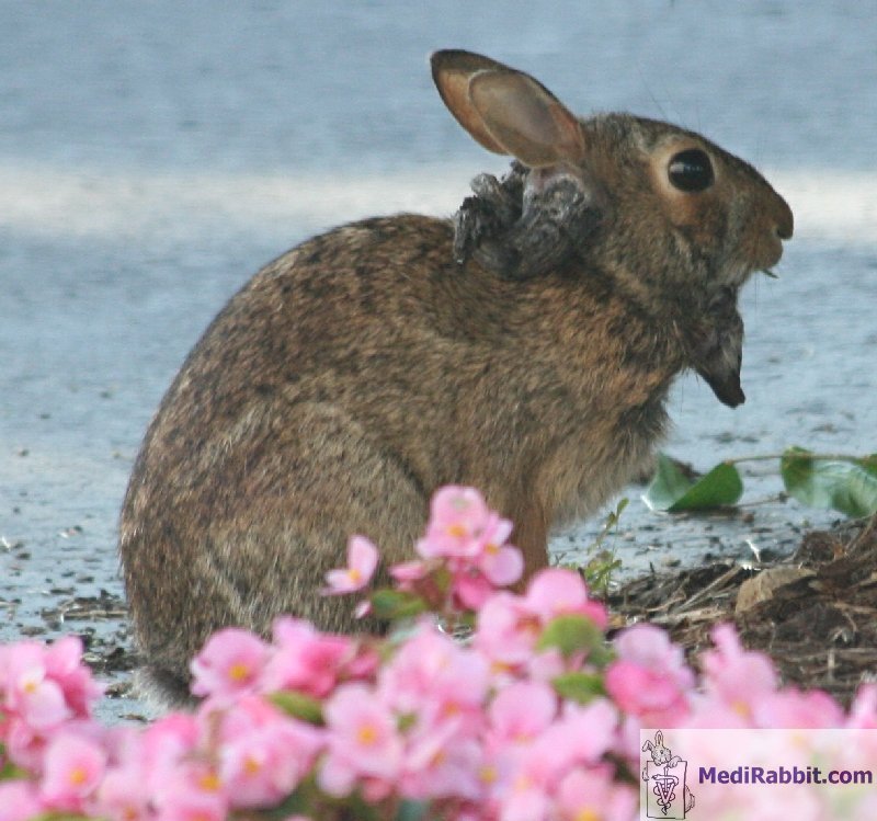
Between
the 16th and the 18th century, illustrations show the
legendary horned rabbit, a hypothetical cross between the antelope and the
hare. Several naturalists studied the horned hare and gave it the Latin
scientific name of Lepus cornutus. It was popularly called “raurackl”,
or "stag-hare”.
It is, nowadays, alleged that the
"horns" around the head of the "Lepus cornatus" do
not relate to imagination, but to the growth of papilloma or fibroma tumors. The latter
develop after infection of a cell with the Shope papilloma virus, Shope
fibroma virus or the leporipoxvirus.
Shope fibroma virus
The
Shope fibroma virus was discovered in 1931 by R.E. Shope. It is found mainly
on the US continent, among the cottontail (Sylvilagus floridanus)
population. It was soon realized that the virus is transmissible between
cottontails and rabbits. A viral infection results in the development of
gross and microscopic lesions called fibromas.
The virus is spread through bites
of blood seeking insect (e.g. fleas, mosquitoes). Once a cutaneous cell is
infested, it will undergo a transformation leading to the formation of a
tumor.
Shope showed by different
experimental techniques that the fibroma virus is related to the myxoma
virus. This property is nowadays exploited by using the live Shope fibroma
virus to vaccinate against myxomatosis.
European hares are known to carry a virus
(Leporipoxvirus) responsible for fibromatous diseases. Rabbits are
susceptible to this virus. Clinical signs include the growth of numerous skin
nodules (up to 2.5 cm in size) on the face, eyelids and around the ears. The
transmission mode of this virus is unknown. Clinical signs and
diagnosis
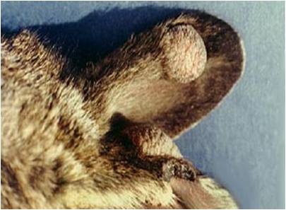
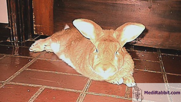
The symptoms of fibromatosis were
accurately described by Shope in 1931, with tumors appearing essentially on
the feet and limbs, and to a lesser extent on the face, the nose, the
eyelids, and the back. In newborn rabbits and cottontails, it causes general
and severe diseases; in adult rabbits, the tumors often regress naturally.
This virus is, furthermore, known to cause a variety of tumors in cattle.
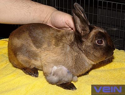
This disease not frequently found
in pet rabbits. The first signs of infection are the thickening of subcutaneous
tissues. A soft well-outlined swelling will grow slowly, and can develop into
a large tumor, with a diameter of 7 cm, and a thickness of 2 cm. The large
size leads to disturbances in the daily activities, such as movement and
search for food.
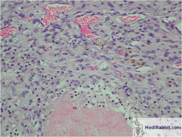
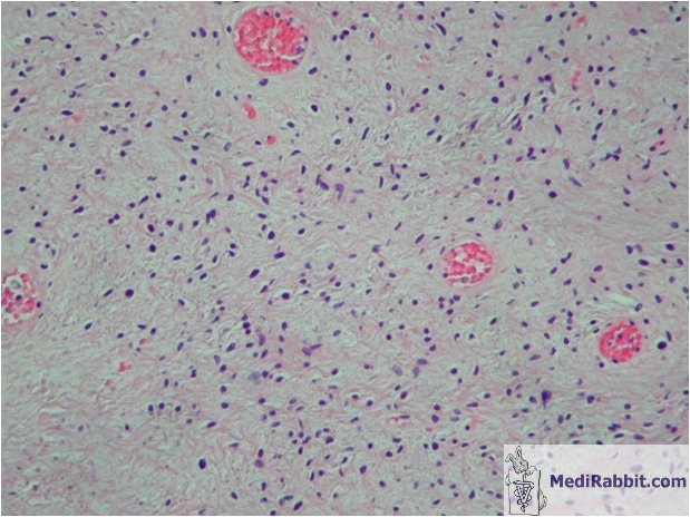
The tumor consists mainly of
connective tissue. Subcutaneous tumors can lead to compression and damage of
the underlying tissues such muscles and tendons. In rare cases, it was
observed that the tumor invades the muscle tissue.
Generally, the tumors regress
spontaneously after 10 to 14 months.
Fibroma tumors must be
differentiated from myxoma and papilloma tumors. The appearance of the
fibroma is usually flat and it is located in the subcutaneous tissues, while
the papilloma tumors have the aspect of a wart, with a well-keratinized
surface.
The diagnosis is based on
clinical signs and can be confirmed with a biopsy sample. Histopathological
examination of the skin lesions shows intracytoplasmic inclusion bodies.
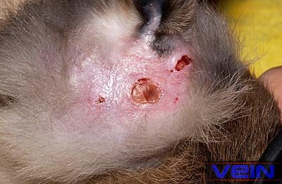
A virus called "malignant
rabbit fibroma virus" has been isolated in rabbits. It can lead to
fibrosarcoma. As it presents antigenic similarities with the fibroma and
myxoma viruses, it is thought to be a recombinant of both viruses, but this
is not yet well defined. The presence of this virus is accompanied by
immunodepression, malignant tumors and infections.
Fibrosarcoma often start in the
soft tissue and spread to a bone by direct invasion or transport of a
metastatic cell via the blood circulation. Secondary fibrosarcoma may develop
in lungs, heart, kidneys and lymph nodes.
Treatment
Surgical
removal is not recommended, because recurrence is quick and usually more
extensive. If excision becomes necessary, it must be wide. For more
details, see: “Fibrosarcoma in rabbits”. For detailed information on fibrosarcoma in
rabbits, by E. van Praag, A. Maurer and T.
Saarony, 408
pages, 2010.
Acknowledgement
Thanks
are due to Jeff Hymel and to Akira Yamanouchi (Veterinary
Exotic Information Network), for
the permission to use their pictures. Further Reading
Hu J, Cladel NM, Pickel MD,
Christensen ND. Amino acid residues in the carboxy-terminal region of
cottontail rabbit papillomavirus E6 influence spontaneous regression of
cutaneous papillomas. J Virol. 2002; 76(23):11801-8. Singh SB, Smith JW, Rawls WE,
Tevethia SS. Demonstration of cytotoxic antibodies in rabbits bearing tumors
induced by Shope fibroma virus. Infect Immun. 1972; 5(3):352-8. Smith JW, Tevethia SS, Levy BM,
Rawls WE. Comparative studies on host responses to Shope fibroma virus in
adult and newborn rabbits. J Natl Cancer Inst. 1973; 50(6):1529-39. Friedman-Kien AE, Fondak AA, Klein
RJ. Phosphonoacetic acid treatment of shope fibroma and vaccinia virus skin
infections in rabbits. J Invest Dermatol. 1976; 66(02):99-102. Block W, Upton C, McFadden G.
Tumorigenic poxviruses: genomic organization of malignant rabbit virus, a
recombinant between Shope fibroma virus and myxoma virus. Virology. 1985;
140(1):113-24. Strayer DS, Cabirac G, Sell S,
Leibowitz JL. Malignant rabbit fibroma virus: observations on the culture and
histopathologic characteristics of a new virus-induced rabbit tumor. J Natl
Cancer Inst. 1983; 71(1):91-104. Strayer DS, Sell S.
Immunohistology of malignant rabbit fibroma virus--a comparative study with
rabbit myxoma virus. J Natl Cancer Inst. 1983; 71(1):105-16. Strayer DS, Skaletsky E, Cabirac
GF, Sharp PA, Corbeil LB, Sell S, Leibowitz JL. Malignant rabbit fibroma
virus causes secondary immunosuppression in rabbits. J Immunol. 1983;
130(1):399-404. Strayer DS,
Skaletsky E, Leibowitz JL, Dombrowski J. Growth of malignant rabbit fibroma
virus in lymphoid cells. Virology.
1987; 58(1):147-57. |
e-mail: info@medirabbit.com
