![]()
Myiasis
(botfly) in rabbits
Esther van Praag Ph.D.
|
|
MediRabbit.com is funded
solely by the generosity of donors. Every
donation, no matter what the size, is appreciated and will aid in the
continuing research of medical care and health of rabbits. Thank you |

Warning: this file
contains pictures and videos that may be distressing for people.
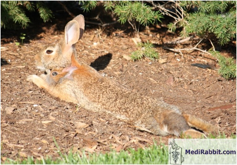
Myiasis caused by larvae of the Cuterebra sp. flies is found only in the USA. It
is most commonly observed during the hot humid summer months and during fall,
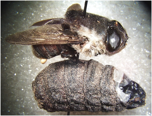
and affects mainly younger animals. Cuterebra
sp. flies are large, hairy, and characterized by the absence of a functional
mouth. Their life span is short, and aimed only at the reproduction of the
species. The larvae of several species of the Cuterebra
sp. flies can infest rabbits and other lagomorphs. They include Cuterebra buccata,
C. cuniculi, C. lepivora, C. abdominalis, C. jelloni,
C. ruficrus, and C. lepusculi.
The parasitic larvae of these flies can infest human beings and other animals
as well, including dogs, foxes, cats, and minks.
Unlike with fly-strike, a Cuterebra sp. larva strike is not linked to poor
hygiene. Indeed, the eggs are not deposited on skin soiled with urine or
excrement, but near the entrance to a rabbit burrow, other lagomorph nests,
or near an outdoor rabbit hutch. House rabbits can also be struck by botfly
larvae, when a fly enters a home, and deposits eggs in the rabbit's living
environment. When the botfly larva emerges from the egg, it will migrate onto
a (wild) rabbit, cottontail, or hare. It enters the body of its host through
the skin (breaks in the skin or any natural openings), after which it
penetrates the mucosa. The larva will migrate further in the body, using the
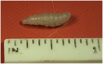
trachea and the abdominal cavity to move to a subcutaneous location. There it
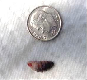
will develop a 2 to 3 cm long furunculoid cystic
structure, with a fistula (respiratory hole) at the surface of the skin, and
swelling of the subcutaneous tissues.
Depending on the species of botfly,
the cysts will develop in different parts of the rabbit's body. Larvae of C.
buccata can infest the entire abdominal region
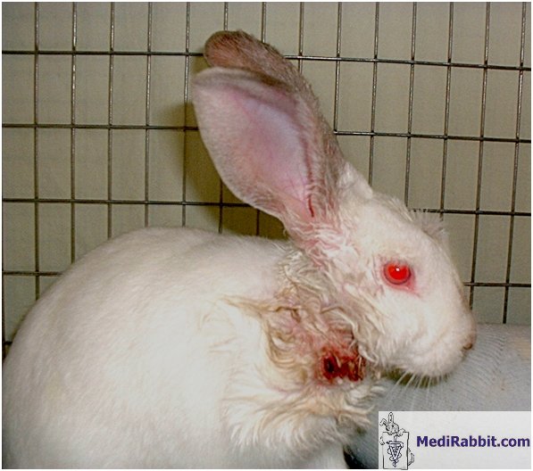
(especially the inguinal area, abdomen or shoulders), whereas larvae of C.
horripilum have mainly been observed in the
cervical region. When the larva
reaches the stage of pupation, it disengages from the cyst and falls off.
Clinical
signs
The
clinical signs are generally sufficient for a proper diagnosis.
The
early stages of myiasis are sub-clinical. With time
however, a rabbit becomes depressed, anorectic, dehydrated and weak, loses
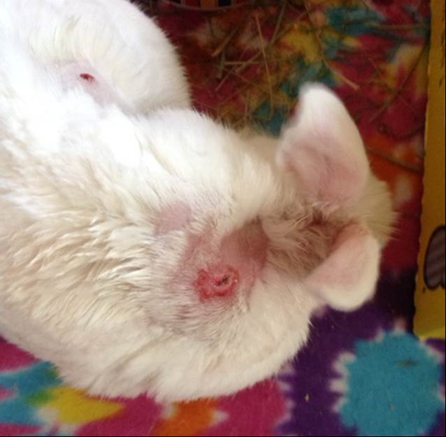
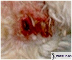
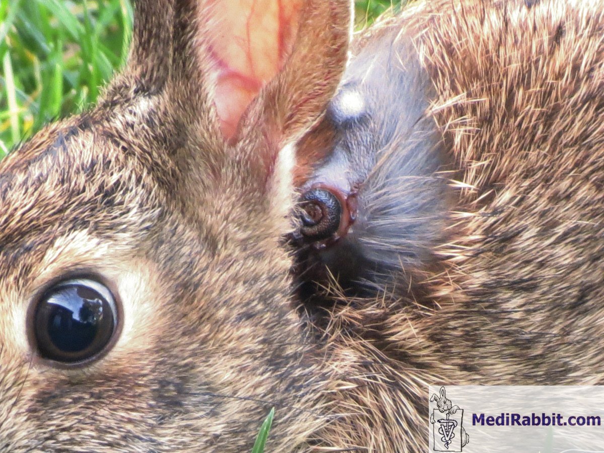
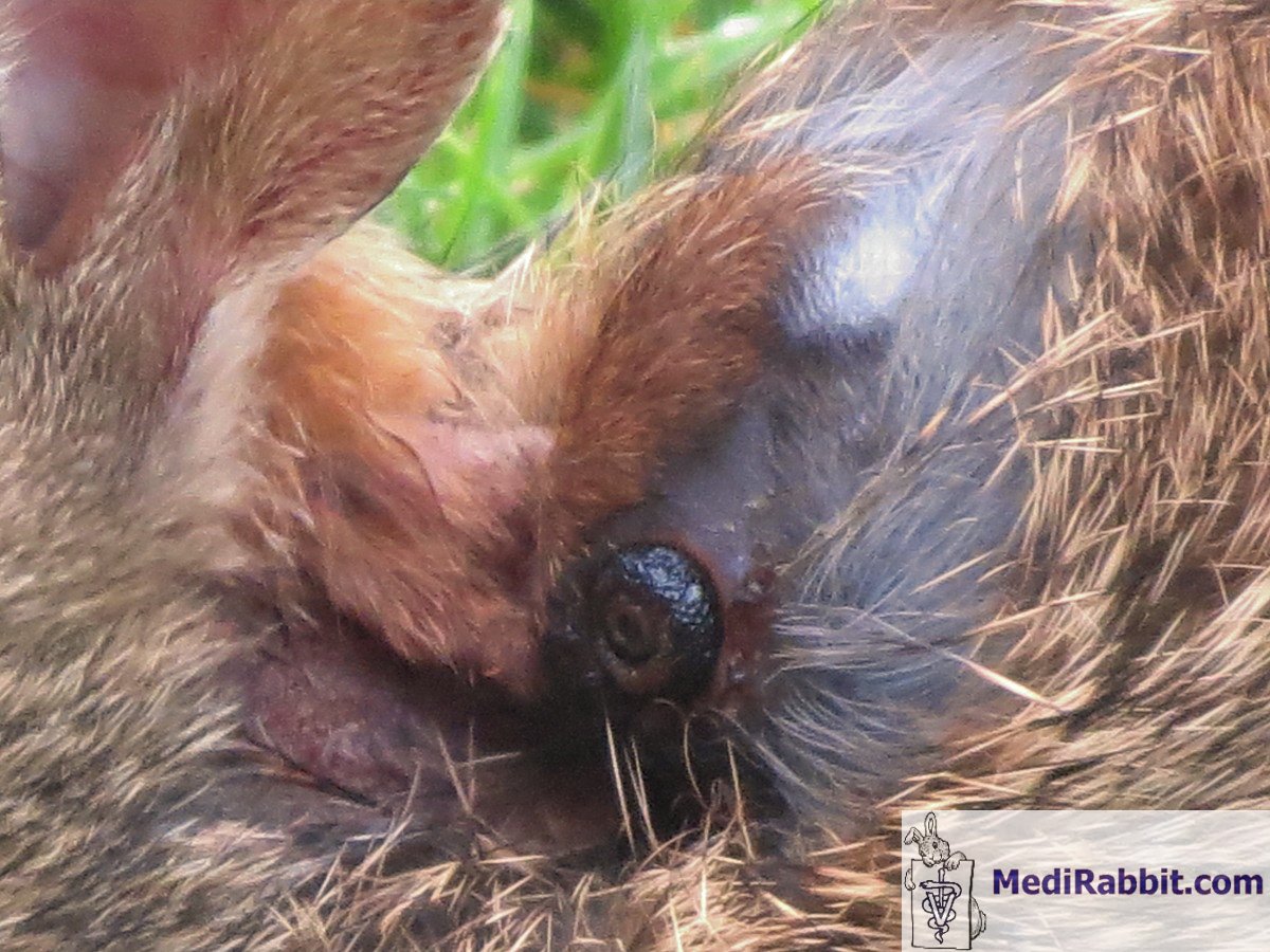
weight, and may go into shock if the infection is severe. At this stage the
infection becomes discernible, with a visible fistula in the skin,
accompanied by a lump or a cystic structure. The lesion is painful, and
causes great distress to the rabbit.
Progressively the skin around the
hole becomes moist, and the surrounding hair matted, leading to the
development of secondary bacterial or fungal infections.
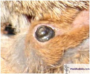
There is potential for aberrant
migration of the larvae into the nasal cavity and sinuses, or the eyes.
Migration into the trachea has also been observed, leading to the formation of
laryngeal edema, blocking the air supply to the lungs, and sometimes
accompanied by concurrent accumulation of mucus, and swelling of the
esophagus. Migration into the brain, via the ear canal is a further potential
danger. Once in the brain, it will cause severe and irreversible neurological
damage.
Diagnosis
The history of the rabbit and the
clinical signs are generally sufficient for a proper diagnosis.
Treatment
The skin is prepared as for a surgical procedure, with the hair
delicately clipped around the infected area, and the skin disinfected with an
antiseptic solution. After enlargement of the breathing hole, the larva is
carefully removed with the aid of forceps, without damaging or crushing it,
in order to prevent skin irritation, and especially in order to prevent the
occurrence of a (fatal) anaphylactic reaction. After removal of the larvae
the cavity is cleaned with a sterile saline solution, an antiseptic solution,
and an insecticide solution. If necrotic tissue is present, the cavity should be carefully
debrided. If an abscess has formed in the cavity, surgical excision of the
tissues is necessary, followed by topical and systemic antibiotic therapy. Aberrant migrated larvae, located deep under the skin or in vital
organs, are removed surgically, under anesthesia. The administration of non-steroidal analgesics (pain medication) is
necessary (e.g. meloxicam, carprofen) after the
procedure. If the affected rabbit stops eating, it should be hand-fed, in order
to avoid fatal hepatic lipidosis. If a rabbit is heavily infested with botfly larvae, euthanasia should
be considered. For detailed information on botfly infestation in
rabbits, see: “Skin Diseases of Rabbits” by E. van Praag, A. Maurer and T.
Saarony, 408 pages, 2010.
Acknowledgement
Thanks are
due to Connie Andrews (USA), to
Joanne Vujnovich (USA), David and Ann Lynch (USA)
and the owner of “Cedar Creek Natural History Area”, and Iris Klimczuk (USA) for the
permission to use their illustrative material. Thanks also to Tal Saarony for
the critical reading of this text.
Further
Readings
Baird CR. Biology of Cuterebra
lepusculi Townsend (Diptera:
Cuterebridae) in cottontail rabbits in Idaho. J Wildl Dis. 1983 Jul;19(3):214-8. Jacobson HA, McGinnes
BS, Catts EP. Bot fly myiasis of the cottontail
rabbit, Sylvilagus floridanus mallurus in Virginia with some biology of the
parasite, Cuterebra buccata.
J Wildl Dis. 1978 Jan;14(1):56-66.
Schumann H, Schuster R, Lange J. The
warble fly Oestromyia leporina
(Diptera, Hypodermatidae)
as a parasite of the wild rabbit (Oryctolagus cuniculus). Angew Parasitol. 1985 Mar;26(1):51-52. Weisbroth SH, Wang R, Sacher S. Cuterebra
buccata: immune response in myiasis
of domestic rabbits. Exp Parasitol. 1973 Aug;34(1):22-31. |
||||||||


e-mail: info@medirabbit.com
