![]()
Fungal
Dermatitis or Superficial Mycosis
Esther van
Praag, Ph.D.
|
MediRabbit.com is
funded solely by the generosity of donors.
Every
donation, no matter what the size, is appreciated and will aid in the continuing
research of medical care and health of rabbits.
Thank you
|

Warning: this file
contains pictures that may be distressing for people.
Mycosis is not common
in either wild or house rabbits and is rarely encountered as epizootic. Rather,
it generally occurs as a sporadic infection in one individual rabbit, in
young rabbits, or in sick debilitated rabbits. It is believed that
immunosuppressed rabbits are more susceptible to the disease.
The causing agents are
two pathogenic fungi:
- Trichophyton
mentagrophytes = ringworm
May be carried asymptomatically in the coat of the rabbit.
- Microsporum canis
Occasionally rabbits
are infected through contact with cats or dogs; this fungus can carry one or
more zoonotic diseases (diseases that can be transmitted from animal to
human).
Ringworm is the main
cause of mycosis. It will infect not
only the epidermis, but also the annex structures such as hair follicles and
hair shafts. In some cases, cultures of infected tissue have revealed the
presence of Microsporum sp.
Clinical signs
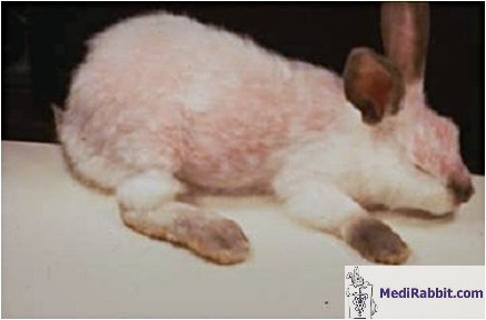
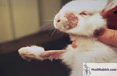
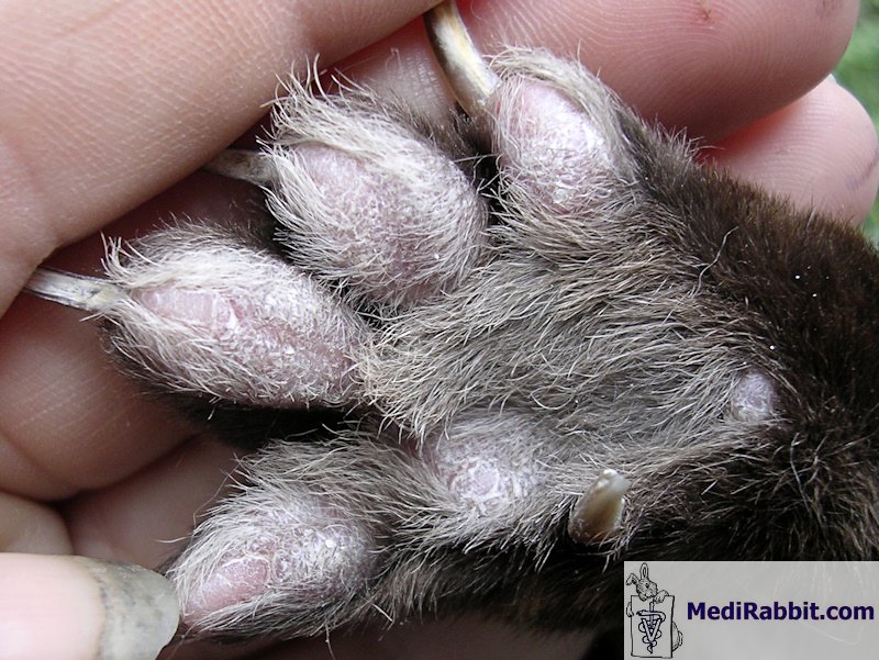
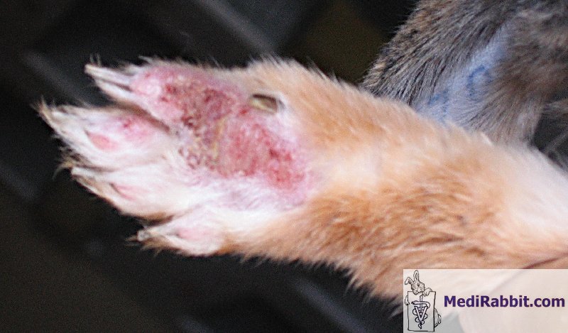
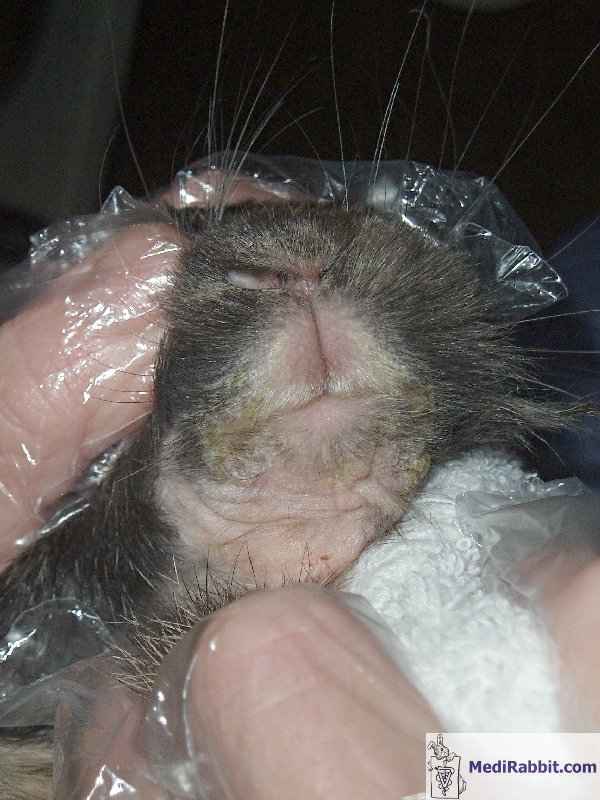
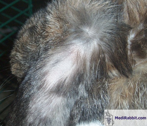
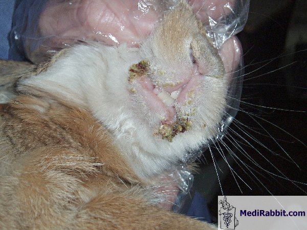
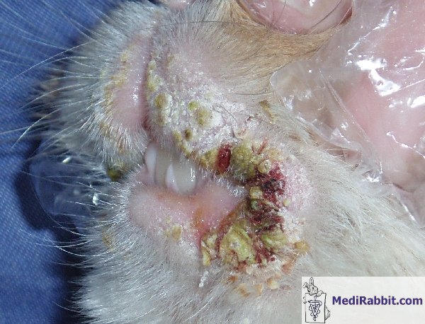
Typically, lesions
start around the head and spread to the legs and feet, more specifically to
the toenail beds. The wound is raised, circumscribed and erythematous. It
shows dry crusts with little or no pruritus and patchy alopecia. The tissue
under the crusts usually shows inflammation and the hair follicles show
abscessation, as the secondary result of a bacterial invasion.
Histological sections show
hyperkeratosis, folliculitis, acanthosis and the diffuse infiltration of
leukocytes into the dermis layer.
Diagnosis
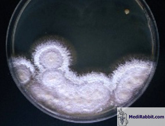
1. Cultures on a fungal or dermatophyte
media. It must, however, be kept in mind that the results obtained may not be
reliable and must be completed with histological studies.
2. Identification of skin scrapings mounted
in 10% KOH. This enables to identify the different arthrospores
3. Fluorescence (UV light) is of little
help. One fungus (Trichophyton mentagrophytes) does not fluorescence; the
other fungus (Microsporum
canis)
is strain dependent: some show fluorescence, others do not.
4. Gomori methenamine silver stain, Gridley fungus
stain and periodic acid-Schiff (PAS) reaction staining are all methods that
help demonstrate the presence of arthrospores and to identify them.
The
diagnosis must differentiate from other causes of crusty alopecia commonly
found on the head and ears (genetic hairlessness, trauma, depilatory hair
loss, fur pulling).
Treatment
The hair around the
lesion should be clipped, and disposed off safely.
The best treatment for
fungal dermatitis is oral administration of griseofulvin (25-50 mg/kg PO q24h
or divided q12h). (Wear gloves while administering this drug). The treatment
should continue two weeks after the disappearance of the clinical signs.
Topical or systemic
treatments are also possible:
• Clotrimazole cream or lotion;
• Enilconazole spray;
• Itraconazole (5-10 mg/PO, q 24 h);
• Terbinafine (8-20 mg/kg PO, q24h);
• Ketoconazole (10-15 mg/kg PO q 24h) (not to
be used in breeding animals);
• Miconazole cream.
Most of the available products
are unlicensed for use in rabbits; literature references, however, assert
those drugs are the treatments of choice for fungal dermatitis in rabbits.
The
environment of the rabbit should be cleaned carefully: vacuum cleaning,
boiling of towels and cleaning of surfaces with 1:10 bleach water.
Acknowledgement
Thanks are due to Lynne Huntley (USA)
and to Kathleen Hermans, D.V.M. (Kliniek voor Pluimvee en Bijzondere Dieren, Universiteit Gent, Belgium) and
to Prof. Richard Hoop (Institut für Veterinärbakteriologie, University of
Zurich, Suisse) and to Michel Gruaz (Switzerland) for their pictures.
Further information
K. L. Banks, T. B. Clarckson (1967) Naturally occurring dermatomycosis in the rabbit. J. Am.
Med. Assoc. 151: 926-29.
K. W. Hagen (1969) Ringworm in domestic rabbits:
Oral treatment with griseofulvin. Lab Anim. Care 19: 635-638.
E. V. Hillyer, K. Quesenberry, S. Valkoff (1997)
Ferrets, Rabbits and Rodents: Clinical Medicine and Surgery, (Editor)
Saunders W B Co, p. 215.
L. M. Vogstberger et
al. (1986) Spontaneous dermatomycosis due to Microsporum canis in
rabbits. Lab. Anim. Sci. 36: 294-97.
|
|||||||||||||||||

e-mail: info@medirabbit.com
