![]()
Case report:
Atypical treponematosis - rabbit syphilis
in a pet rabbit
Tal Saarony
|
|
MediRabbit.com is
funded solely by the generosity of donors. Every
donation, no matter what the size, is appreciated and will aid in the
continuing research of medical care and health of rabbits. Thank you |

Warning: this
file contains pictures that may be distressing to some persons
|
Summary
about the general form of rabbit syphilis The 2
most important facts about rabbit syphilis are: 1.
That it is completely treatable and curable; 2.
That it is not zoonotic and cannot be transmitted to
humans. Rabbit syphilis is a disease caused by the bacterium
Treponema cuniculi. It can be transmitted sexually, but has also been seen
in rabbits that were living singly, having had no contact with other rabbits,
and in rabbits that were sharing space with unaffected rabbits. It is
believed that the disease was transmitted to these rabbits at birth or via
the mother's milk while nursing. In some rabbits the bacterium may remain
dormant for long periods of time, even years, and the affected rabbit will
show no clinical signs until a stressful event occurs,
causing the infection to erupt. The incidence of syphilis in house rabbits is not
known, but it is likely more common than previously thought. It is,
therefore, important to be aware of the clinical characteristics of the
regular and atypical forms as well as of the easy availability of treatment. Clinical signs The typical form of syphilis affects the mucocutaneous junctions of the genitalia, the anus and/or
the face, mainly around and on the eyelids and nose. Lesions develop slowly.
The skin becomes crusty and ulcerated. The secretion of a pus-like
exudate and bleeding can occur. Due to slow immune response to the bacterium,
the infection can spread to
the surrounding areas and other
susceptible regions on the body (e.g., from the anus/genitalia to
the face) when left untreated. Over the last years, an atypical form of
treponematosis has been observed in rabbits, in which clinical signs are seen
only on the face and not on the genitals/anus. The affected area will exhibit
lesions that will develop into crusts and, if left untreated, will spread.
The lesions may become raw, inflamed, or may bleed or exude a discharge. Treatment Syphilis bacteria will only be killed with injected
penicillin G, at intervals of 5 to 7 days, continued 4 - 5 weeks. The
importance of injected administration must be emphasized here: rabbits should
NEVER be administered penicillin orally. It is important to monitor the rabbit's eating. As
the dying bacteria release toxins inside the rabbit's body, the appetite may
be affected. Inappetence can last 2 - 3 days, but the rabbit usually begins
eating again on its own. It is essential that the antibiotic is not stopped;
the rabbit should be encouraged to eat, tempted with favorite foods, and
given a lot of attention and love to motivate it to eat. References 1.
Saito K., Tagawa
M., and Hasegawa A. Rabbit Syphilis Diagnosed Clinically in Household Rabbits
J. Vet. Med. Sci. 65(5): 637-639, 2003 2.
Saito K., Tagawa
M., and Hasegawa A. RPR Test for Serological Survey of Rabbit Syphilis in
Companion Rabbits J. Vet. Med. Sci. 65(7): 797-799, 2003 3.
Bellangeon M. Tréponématose chez le
Lapin de Compagnie PASE 2, deuxième trimestre 2001 4.
Saunders RA, Davies RR. Notes of
rabbit internal medicine. Oxford, UK: Blackwell Publishing; 2005 5.
Quesenberry KE, Carpenter JW.
Ferrets, rabbits and rodents. Clinical Medicine and Surgery. St Louis, USA:
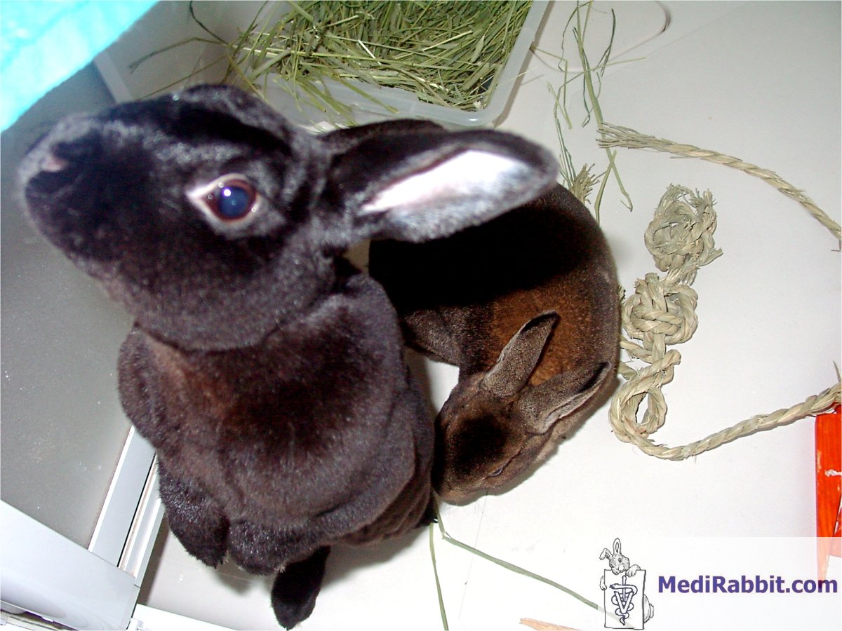
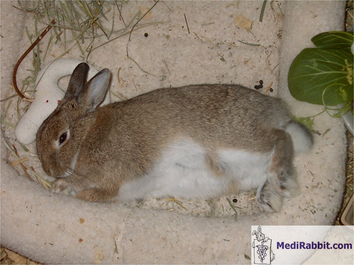
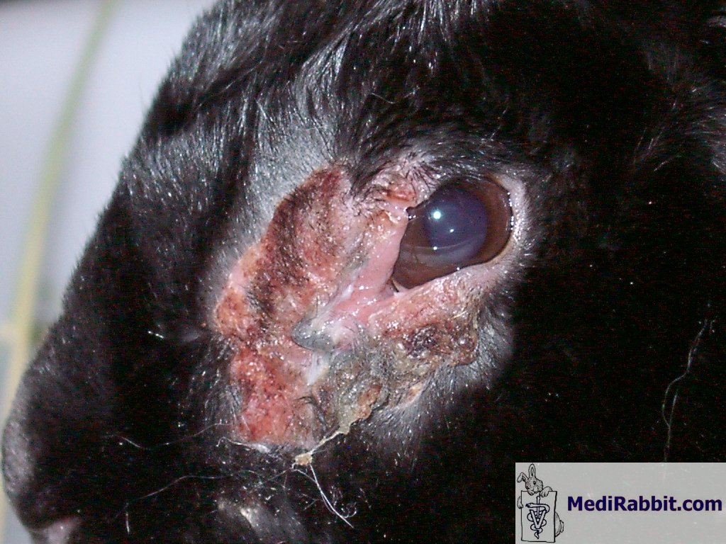
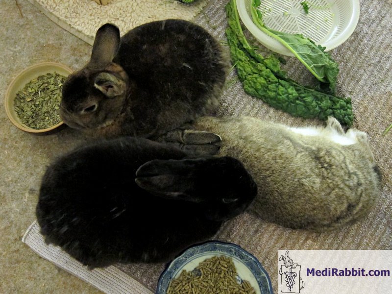
Saunders; 2004 Gozal's syphilis The cast of characters:
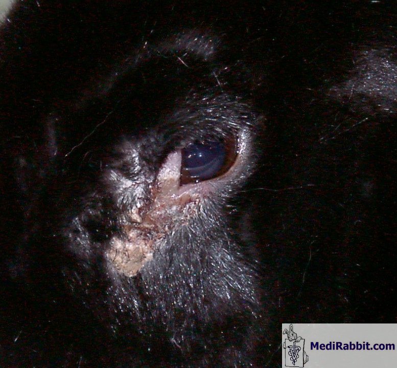
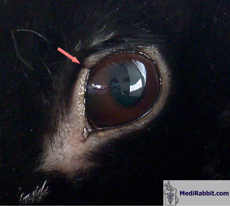
On Feb. 9th, 2007 I first discovered what appeared
to be an injury at the corner of Gozalʼs eye. I believed it resulted from
an altercation during a bonding session with Tinok. It resembled a crusty eye discharge.
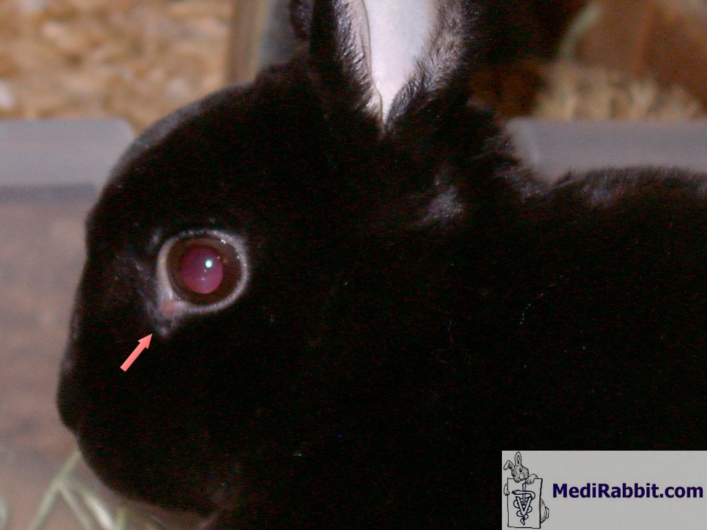
Later it began to appear inflamed. The following picture was taken on
March 5th:
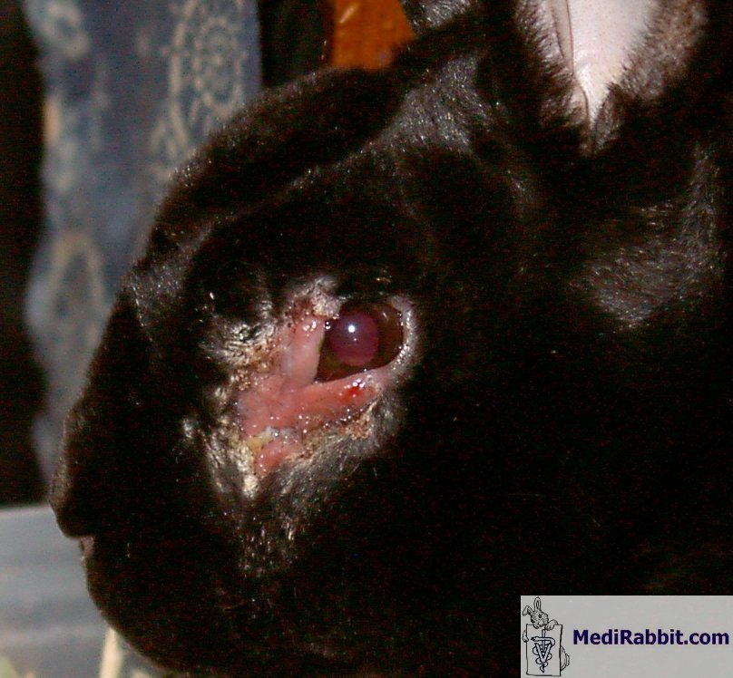
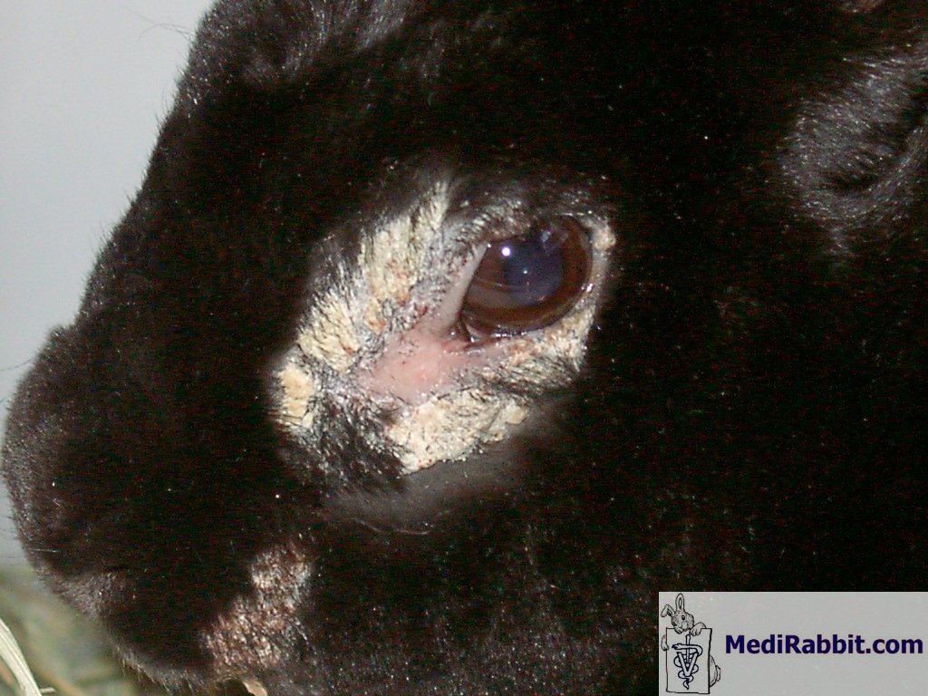
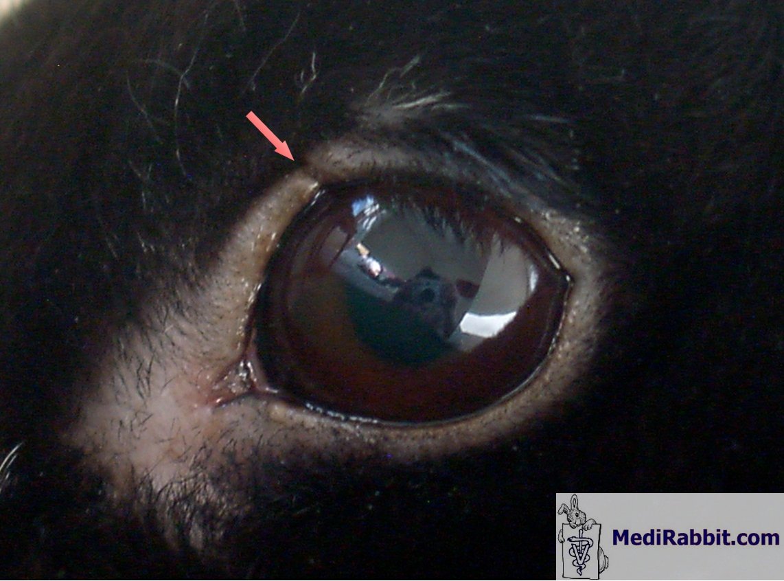
Motek was grooming the eye excessively and it was believed
she was not allowing the wound to heal. The wound was crusted over, then,
suddenly, a crater appeared (the ulceration that formed underneath the crust,
± 1cm into the skin). It was not clear whether the crust fell off
spontaneously or was pried off by Motek. They were separated. Gozal was being
treated with an antibiotic eye ointment which had no effect on his condition.
The wound also began bleeding. April
18th:
April
20th:
May
8th:
May10th:
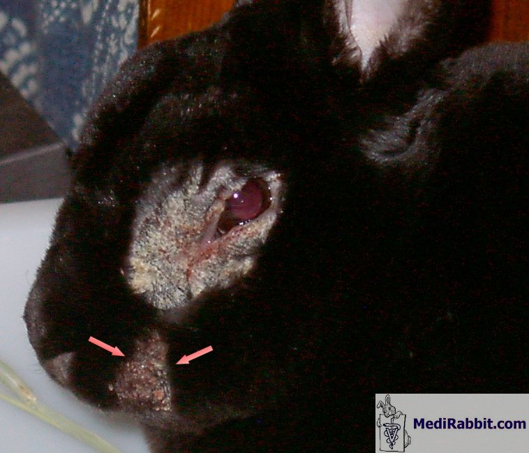
Around that time Gozalʼs nose presented with crusts as well,
though to a much
lesser degree; I did not manage to take clear pictures. The crusts on his
face extended approximately 1 cm deep into the skin.
Gozal was injected with a vitamin solution (B1,
B2, B6, B12, A, D, and E), and with the antibiotic
sulfamonomethoxine; Panolog,
an ointment used for external ear infections and skin disorders, was applied
to his crusts. The Panolog application was to be
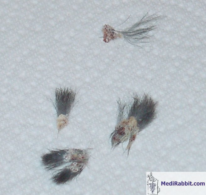
continued at home. Gozal’s wound was debrided by
his vet and the sample crusts inspected under the microscope. Nothing specific was found. A fungal culture was
taken and sent to a lab. No fungi were found. The skin
condition continued to spread. New crusts appeared. May 13th:
May 15th vet visit: Gozal appeared to be responding
to the medication. His tear ducts were flushed under anesthesia and his teeth
checked for abnormal root growth that might affect the eye. The vet
considered a nasolachrymal canal infection (infection
of the canal containing the tear ducts). Gozal was injected with the same
vitamin solution as before and with the antibiotic chloramphenicol, and more Panolog was applied. At that point no diagnosis had yet
been arrived at. I was to continue applying the Panalog
at home and to monitor Gozal for appetite, output, behavior, and the
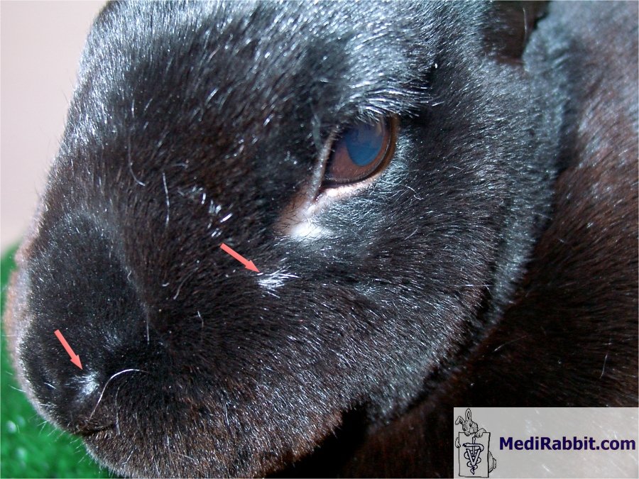
condition of the skin. May
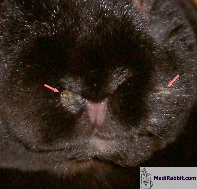
16th:
Crusts on the nose (arrows):
May 22nd: The vet was satisfied with Gozal’s
progress; he observed new hair growth. He applied more ointment and dripped a
fluid to protect from further infection into the eye. More vitamins were
injected as well as chloramphenicol. May
29th: The vet felt Gozal’s condition
had worsened. The crusts had spread. He collected a skin scraping sample, but
was not able to identify anything under the microscope. He ruled out a fungal
infection. Gozal’s tear ducts were flushed under
anesthesia once again. Gozal was injected with Baytril
(enrofloxacin) and a fluoroquinolone preparation (an antibiotic) was applied
to the affected areas. The wound was cultured and the sample sent to a lab
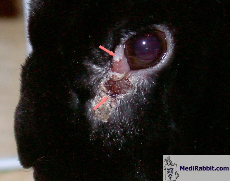
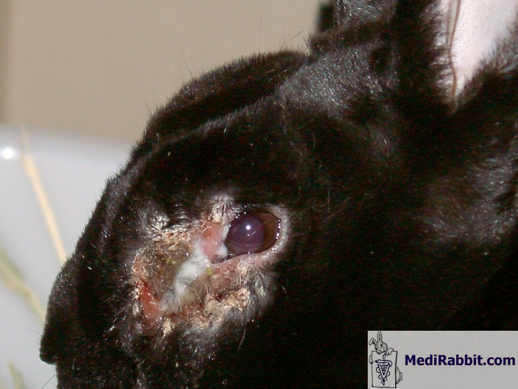
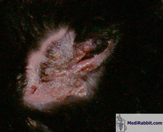
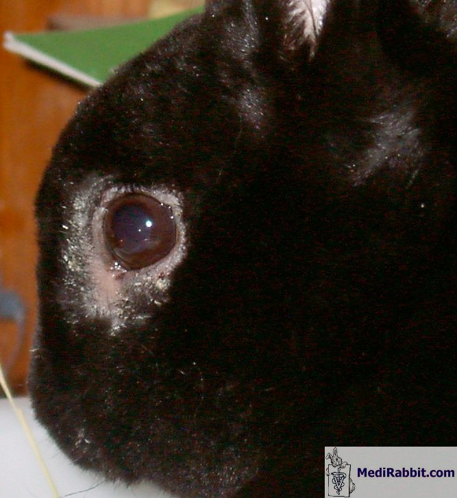
for analysis. May 31st, the lesions/crusts have
spread downward:
Around this time, a friend who is a vet with great
interest in rabbits, and who had been following Gozalʼs condition via emails and
pictures, suggested rabbit syphilis. The information with articles and treatment options
was brought to Gozalʼs vet
who, at first,
dismissed it. However, as Gozalʼs condition was not improving with the administered treatments and all
tests were returning inconclusive, he agreed to try treating Gozal for the
disease. In retrospect this explains why remission was observed after each
administration of antibiotics (see: Treponematosis
– rabbit syphilis). On June 1st Gozal got the first shot of
penicillin. June 4th, immediate, great
improvement:
June
9th:
July
7th:
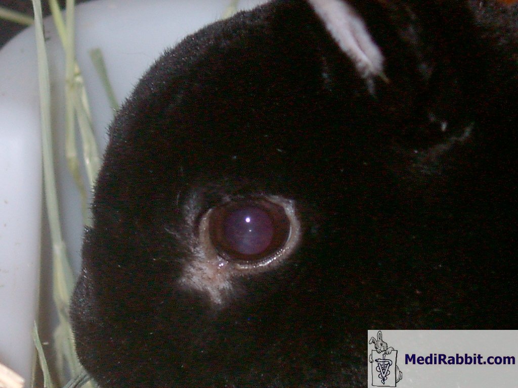
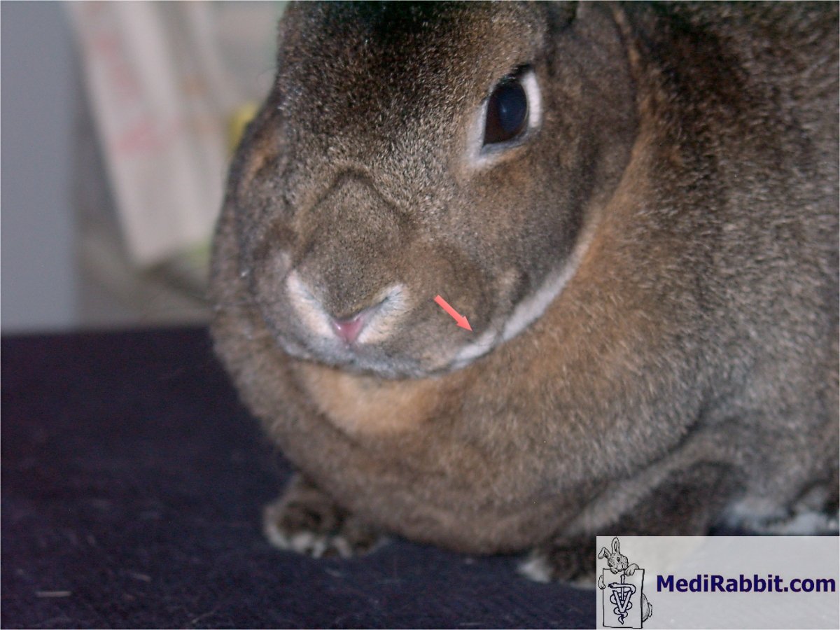
Healed! There is
still a small area at the corner of the eye where hair doesnʼt grow and a cut on the eyelid
where the skin did not regenerate fully (arrows), but Gozal is 100% healthy.
In other spots of previous deep skin lesions the hair grew back white.
Gozal received a total of 5 injections of penicillin
G, a week apart, during 5 weeks. After the first shot he experienced loss of
appetite for about 36 hours, but resumed eating on his own. During his
illness he never stopped eating and never showed any signs of pain or
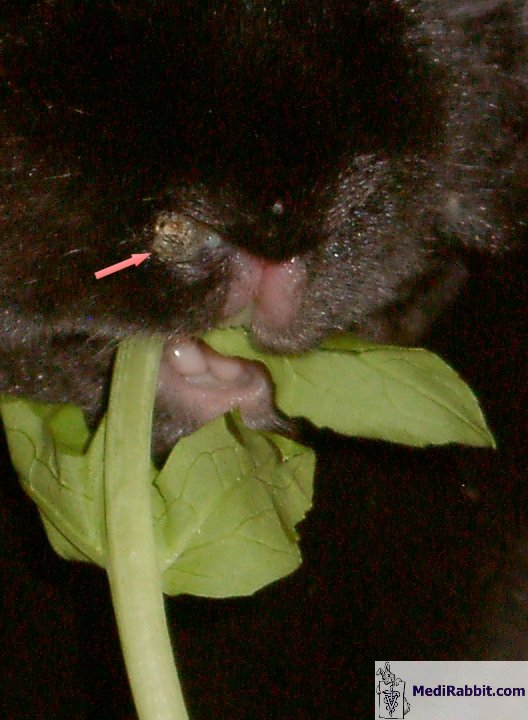
discomfort. Tinok was not infected; 4 months after first
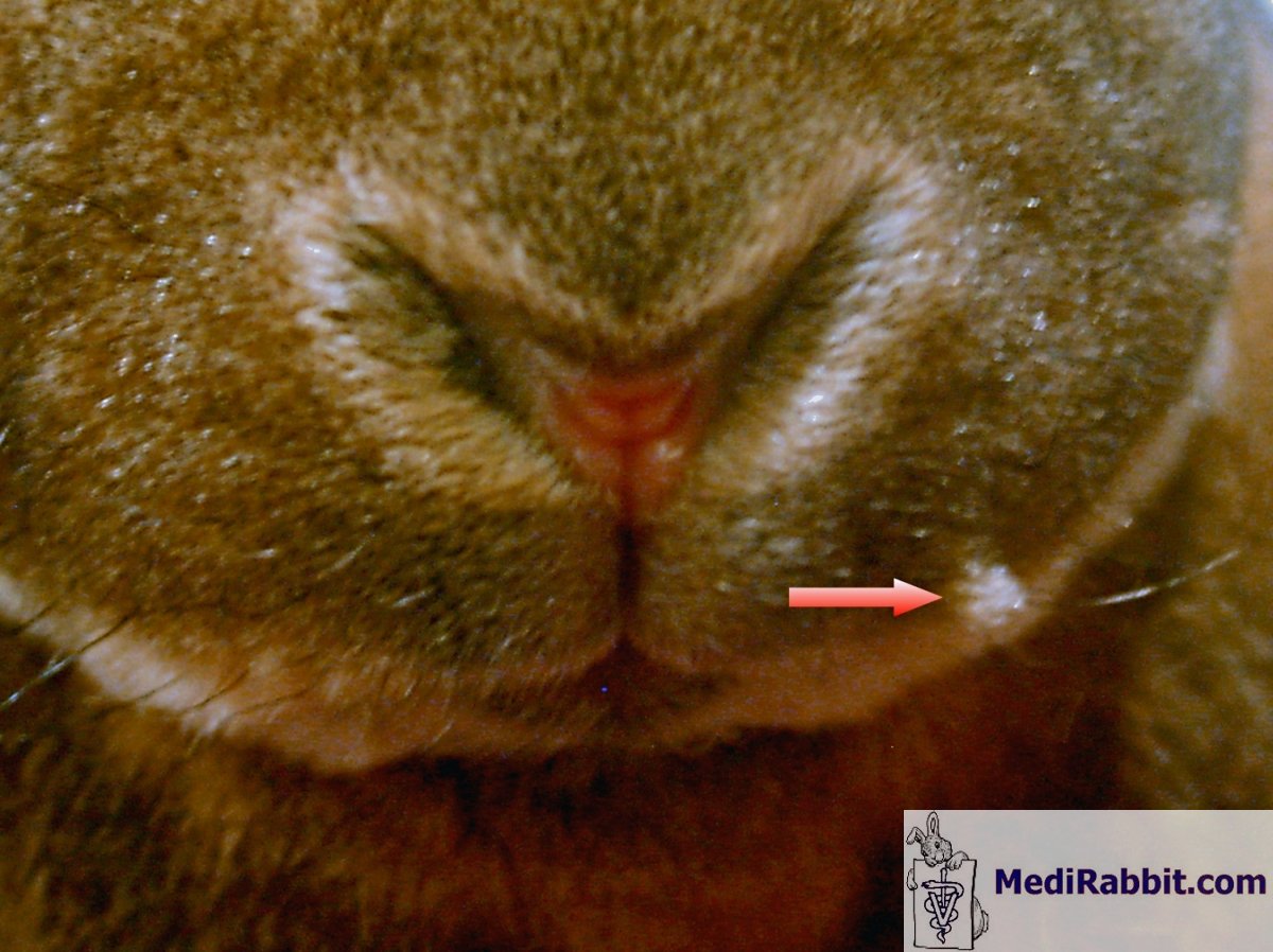
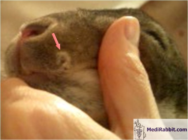
discovering Gozal’s eye, I noticed that Motek’s nose looked very pink and there was a small
protuberance of what appeared to be a raised tuft of hair on the left side of
her face.
The protuberance was softish,
with clumped hair covering it. It was not crusty. While the lump did not look
or feel like the crusts Gozal had exhibited, the pink nose did appear similar.
The vet concluded the lump was the same as Gozal’s
and diagnosed her with syphilis as well; however, the diagnosis was based
strictly on a visual examination and on her proximity to Gozal and previous
extensive grooming of his infected skin. No tests were performed on her. She
received 2 pen G injections, a week apart. Following the first injection the
lump disappeared and the nose improved. She was back to normal after the second injection.
Since Gozal had been exclusively with Motek and I
for a year and a half, and a bit with Tinok, and was already over 3 years old
before showing any clinical signs, it is believed that the disease passed to
him from his mother at birth or while nursing on her milk. It lay dormant and
asymptomatic and most likely erupted due to the stress of bonding. Gozal
dislikes Tinok and to this day they are not bonded. At the time I was
conducting daily bonding sessions. Of my 3 Gozal is the most sensitive and
prone to stress, and his body and immune system reacted by developing the
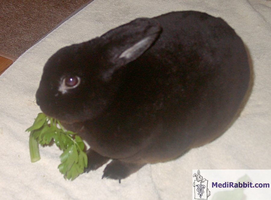
disease. Gozal,
September 18th, 2009.
|
|||||||||||||||||

Many thanks to Motek and Gozal for their patience and their cooperation in
taking the numerous pictures, and to Tinok.
e-mail: info@medirabbit.com
