![]()
Skin Abscesses
Esther van Praag Ph.D.
|
MediRabbit.com is
funded solely by the generosity of donors.
Every
donation, no matter what the size, is appreciated and will aid in the continuing
research of medical care and health of rabbits.
Thank you
|

Warning: this page contains pictures that may be distressing for some persons.
|
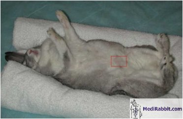
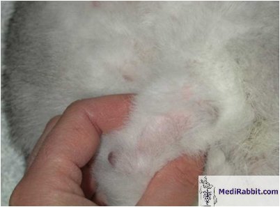
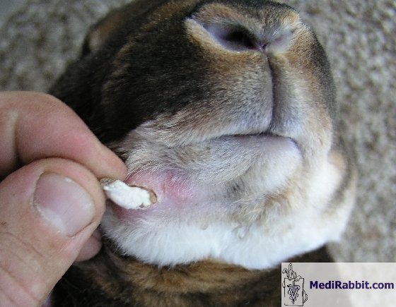
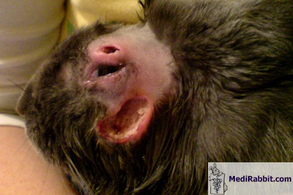
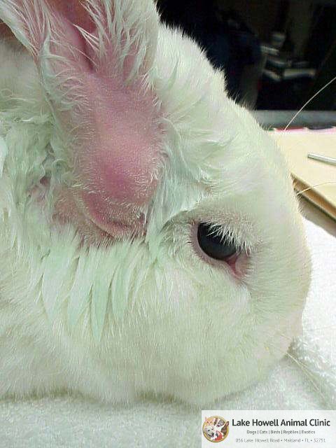
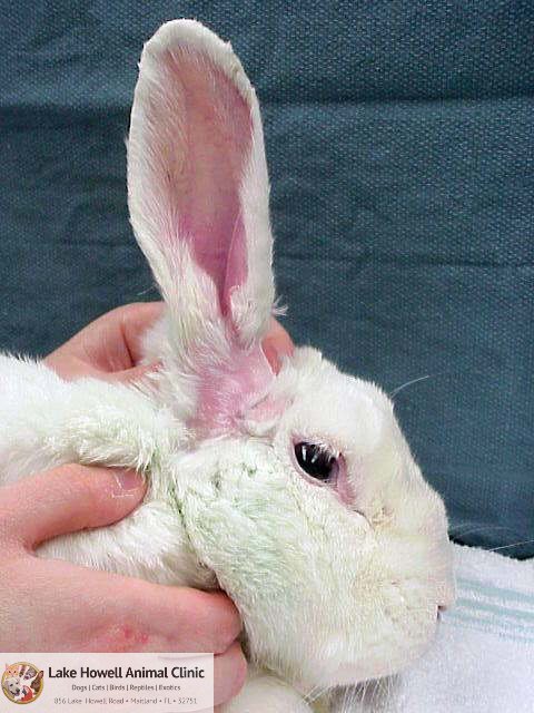
An abscess is a pocket of fluid and pus, which results from an attack by pyogenic organism (e.g., bacterium) followed by the destruction of cells. The pocket usually contains a collection of pus, dead phagocytic white blood cells, necrotic cells, and live or dead bacteria. Pus is particularly thick in rabbit; their heterophil cells (rabbit equivalent of neutrophils) contain a very low level of the enzyme myeloperoxidase, as compared in other animals (dogs, cats) or man. As a result, digestion and liquefication of the material contained in the abscess is slow and pus remains thick and sticky. As the quantity of pus increases, the pocket grows larger and starts to wall off from the surrounding tissues and blood circulation. This renders treatment difficult. If the abscess is left untreated, it continues to grow. Tissue will rupture either inside the body or on the surface of the skin. This stage is dangerous, with the liberation of bacteria and their toxins in the blood circulation.
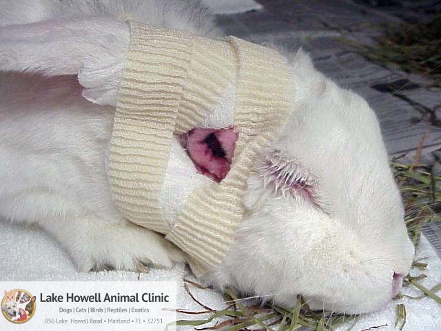
Caroline Charland
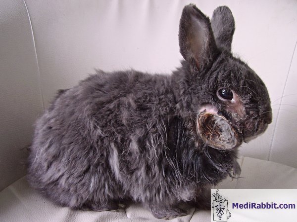
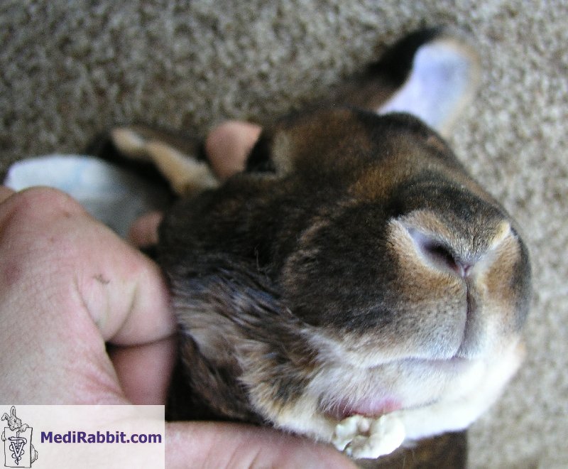
Rescued rabbit with a runny eye and a facial abscess that was left untreated. As a result, it burst and dried out. The presence of the abscess, the pain and poor health affected the quality of the fur, which look dull and unkept In most cases
of abscess, the rabbit has a history of pasteurellosis,
though other bacteria like Staphylococcus aureus, Streptococcus
sp., Pseudomonas sp or Fusiformis sp.
are no exceptions. They often result from a nasolacrimal or tooth problem,
surgery, insect bites, scratches or skin wound, trauma, or a foreign body
such as a hay splinter in the gum, foot sole or anal gland. Abscesses are
typically found on the head, neck and shoulders regions, but can also affect
other parts of the body, organs or bones. During palpation, abscesses feel
either as a hard lump or a soft doughy swelling that can be moved. The
presence of an abscess is usually painless, unlike other animals. It is often
observed that abscesses can grow within a few days time.
Clinical signs
There are no specific
clinical signs, except the presence of a lump, an unusual swelling, reduced
appetite, increased water intake, and/or fever.
Treatment
The treatment
of an abscess is difficult, long, and recurrence is no exception in rabbits. Use
of systemic antibiotics will have little effect and more aggressive therapy
is needed. The best
option remains total surgical excision of the abscess cavity, necrotic
tissue, and the surrounding fibrous capsule. This treatment can, however, not
be done when several abscesses are present, or when the bone is affected
(e.g., osteomyelitis, jaw bone infection by a tooth root). During the
surgical procedure, it is important to check that no fibrous channels leading
to abscess cavities deeper in the tissue remain. If present, the cavities
must be flushed with antiseptic solutions (e.g., chlorhexiderm
or iodine povidone) by means of a catheter tube. A drain can be placed to
facilitate this procedure. The cavity
should be packed with an antibiotic impregnated dressing. Various types are
available nowadays include: • Permanent placement of antibiotic
impregnated PMMA beads; • Temporary filling with antibiotic
impregnated gelatin or cellulose hemostat (e.g., GelFoam®,
Surgicel®). The dressing must be changed daily or
every 2nd day, to avoid necrosis of surrounding tissues; • Temporary filling with wet-to-dry
hygroscopic and bactericidal sugar dressing (e.g., 50% dextrose, or manuka or clear sterilized (g-rayed)
honey). The dressing must be changed daily, to avoid necrosis of surrounding
tissues. The later
filling presents the advantage to remove the malodorous smell of ammonium and
sulfur compounds due to bacterial breakdown of serum or cell proteins. If
osteomyelitis (bone infection) has developed, systemic administration of
antibiotics is necessary. Antibiotics of choice include those that penetrate
the bone or joints, e.g., chloramphenicol, fluoroquinolone (enrofloxacin or marbofloxacin), bicillin (a
combination of penicillin G benzathine and penicillin G procaine), or
penicillinase-resistant semi-synthetic penicillin such as cephalosporin or
metronidazole. The choice of antibiotics safe for use in rabbit is limited.
The treatment
needs to be aggressive and long (4 to 6 weeks). If this antibiotic treatment
is not successful, or if large parts of the bone have been destructed,
radical debridement remains necessary. Nowadays, gentamycin PMMA beads can be
implanted near the bone. There
is little information is available about the effusion rate of the antibiotic
from antibiotic impregnated gel sponge or beads. Newly grown tissue may
hinder a good distribution of the antibiotics from beads after 3 weeks
already, limiting their action to 6 weeks. It
should be kept in mind that abscesses in rabbits are difficult to treat and a
full recovery without relapse cannot be guaranteed.
Acknowledgement
Thanks are due
(listed alphabetically) to Caroline Charland (www.bunnybunch.org,
USA), to Dr. Orlando Diaz (Lake Howell Animal Clinic, Maitland-FL,
USA), and to Michel Gruaz (Switzerland)
for the gracious permission to use their pictures. Further information
Aoyama T, Sunakawa K,
Iwata S, Takeuchi Y, Fujii R. Efficacy of
short-term treatment of pertussis with clarithromycin and azithromycin. J Pediatr. 1996; 129(5):761-4. Baggiolini M, Hirsch JG, De Duve C. Resolution of granules from rabbit heterophil leukocytes into distinct populations by zonal sedimentation. J Cell Biol. 1969 Feb;40(2):529-41. Blackwell NJ. Abscesses in rabbits. Vet Rec. 1999;
144(19):540.
Harcourt-Brown F. Rabbit Medicine and Surgery,
Oxford, UK: Butterworth-Heinemann 2001, 192 pages. Hillyer EV, Quesenberry QE.
Ferrets, Rabbits, and Rodents: Clinical Medicine and Surgery New York: WB
Saunders Co.,1997. Ladefoged O. The absorption half-life, volume of
distribution and elimination half-life of trimethoprim after peroral administration to febrile rabbits. Zentralbl Veterinarmed A. 1979;
26(7):580-6. Ladefoged O. Pharmacokinetics of trimethoprim (TMP) in normal
and febrile rabbits. Acta Pharmacol
Toxicol (Copenh). 1977;
41(5):507-14. Bergman A, Yanai J, Weiss
J, Bell D, David MP. Acceleration of wound healing by topical application of
honey. An animal model. Am J Surg. 1983; 145(3):374-6. [No authors listed] Related Extraction and abscess
treatment in a rabbit. J Vet Dent. 2000; 17(2):95. Richardson V. Rabbits: Health, Husbandry and
Disease. Blackwell Science Inc,
2000. |
||||||||||||||
e-mail: info@medirabbit.com
