|
Small
Mammal Hematology: Leukocyte Identification in Rabbits and Guinea Pigs
Vanessa
K. Lester, DVM; Heather L. Tarpley, DVM; Kenneth S. Latimer, DVM, PhD
Class
of 2005 (Lester) and Department of Pathology (Tarpley, Latimer) College
of Veterinary Medicine, University of Georgia, Athens, GA 30602-7388
General
Information
Rabbits
and guinea pigs are common companion and laboratory animals. This web
page is designed as a descriptive and visual guide to help
veterinarians recognize the different leukocyte types in
Romanowsky-stained blood smears. Also presented is basic information
concerning leukogram characteristics in these animals during health and
disease.
Most
of the leukocytes in rabbits and guinea pigs appear very similar to
those of other mammals. Novices may easily mistake heterophils (the
lapine and cavian equivalent of neutrophils) for eosinophils.
Heterophils have the same function as other mammalian neutrophils, but
they have acidophilic or eosinophilic granules in their cytoplasm. They
are sometimes referred to as "pseudoeosinophils" in the
literature. Heterophils are present in a number of animal species
including birds, reptiles, amphibians, some fish, rabbits, guinea pigs,
and hamsters. The functions of the leukocytes in rabbits and guinea
pigs are similar to those of other mammals.
Foa-Kurloff
cells are a leukocyte type that is unique to guinea pigs. These are
specialized mononuclear cells that contain an intracytoplasmic
inclusion body of mucopolysaccharide. Although Foa-Kurloff cells can be
present in the blood of both males and females, they are most commonly
observed in blood smears from pregnant guinea pigs. These cells may be
more prominent during pregnancy because they shift from the lungs and
spleen to the thymus and placenta under estrogen stimulation.1
Foa-Kurloff cells possibly function as natural killer cells2.
Leukocytes
of Rabbits
Lymphocyte
- The
morphology of lapine lymphocytes is similar to that of other species.
Lymphocytes have a large nucleus, that may be slightly indented, and a
small amount of light blue cytoplasm. Although small lymphocytes
predominate, large lymphocytes may be present. These cells are similar
in size to heterophils (or neutrophils of other mammals). Large
lymphocytes may occasionally contain azurophilic granules near the
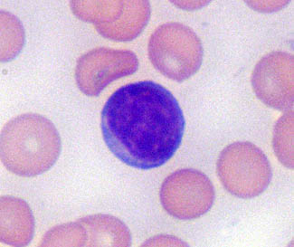
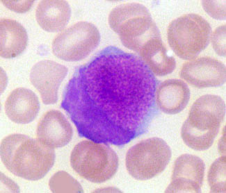
nuclear indentation. Reactive lymphocytes (immunocytes) are
antigenically-stimulated lymphocytes that are larger cells with a more
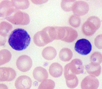
intensely blue cytoplasm (Fig. 1).
|
|
|
|
Figure
1. A normal, small, well
differentiated lymphocyte is on the left, while a larger, reactive lymphocyte
with dark blue cytoplasm is on the right.
|
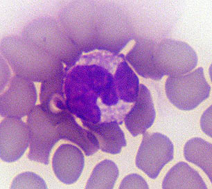
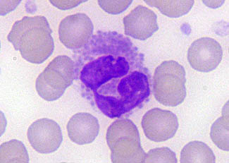
Heterophil
- Inexperienced
microscopists often misidentify rabbit heterophils as eosinophils.
Heterophils range from 10 to15 µm in diameter. They have a light
purple, lobulated nucleus surrounded by cytoplasm containing diffuse,
variably-sized reddish granules. Heterophilic granules are generally
smaller than those of eosinophils and may not occupy all of the
cytoplasm. Although the nucleus is usually segmented, there may be
infrequent band heterophils in the blood of healthy rabbits. Minor
heterophil degranulation may accompany the use of rapid Romanowsky-type
stains such as Diff-Quick. Stain-induced degraulation is presumed to be
a sequel of short fixation time (5 to 10 seconds) during staining.
Degranulated cells will resemble heterophils, but vacuoles will be
present where the granules previously were located. Stain-induced
degranulation should not be confused with toxic change in which the
cytoplasm has a blue cast.
|
|
|
|
Figure
2. Normal rabbit heterophils
have a lobulated nucleus and small, diffuse, red, cytoplasmic
granules (left). Stain-induced degranulation of heterophils may be
observed in some blood smears following Diff-Quik staining (right).
|
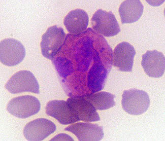
Eosinophil
- Eosinophils
are slightly larger than heterophils and are 12 to 16 µm in diameter.
The nucleus stains purple and often appears bilobed. Intensely
acidophilic, round, cytoplasmic granules are present that are larger
and more numerous than the granules in heterophils.
|
|
|
Figure
3. Normal rabbit eosinophils have
a lobulated nucleus and numerous, round, intensely red, cytoplasmic
granules.
|
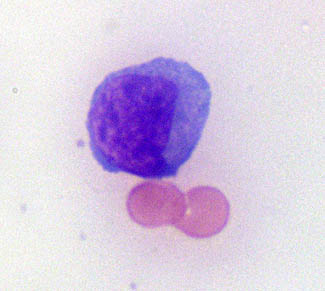
Monocyte
- Monocytes
are the largest circulating leukocytes in health and measure 15 to 18
µm in diameter. Monocytes have a large, variably-shaped nucleus with
chromatin that appears less condensed than that of heterophils. The
cytoplasm is abundant and stains gray to blue-gray. A few cytoplasmic
vacuoles may be observed. Large, dark red granules have been described
in the cytoplasm of some monocytes in association with nonspecific
toxicity.3
|
|
|
Figure
4. A rabbit monocyte with a
nonlobated nucleus and abundant blue-gray cytoplasm.
|
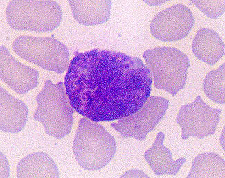
Basophil - Basophils
have a light purple, lobulated nucleus and dark purple to purple-black
cytoplasmic granules. They are approximately the same size as
heterophils.
|
|
|
Figure
5. A rabbit basophil with a
lubulated nucleus and chunky purple granules that partially obscure
nuclear morphology.
|
Comments
Concerning the Hematology of Rabbits
In contrast
to some other mammals, 2 to 4% polychromasia may be a normal
observation in stained blood smears of healthy rabbits. An occasional
nucleated RBC or Howell Jolly body also may be present. The estimated
lifespan of lapine RBCs is 57-67 days.4 This relatively
short erythrocytic lifespan is associated with increased polychromasia
to replace senescent erythrocytes.
Leukocyte
counts are variable both between animals and for different samples from
the same animal. The total leukocyte count is lowest in newborns and
has dual peaks at 3 months and 12 months of age, with a decline between
those time periods.4 There is also diurnal variation in the
leukocyte count with the nadir occurring in the late afternoon to
evening.5 Stress may increase total leukocyte count by 15 to
30%.4
The
relative distribution of rabbit leukocyte subtypes is also variable.
The lymphocyte is the most common leukocyte in the blood of young
animals that are < 12 months of age. After 13 months of age, the
heterophils and lymphocytes may be present in approximately equal
numbers.4 In contrast to many other mammals, healthy rabbits
may have basophils ranging from 5 to 30% of the leukocyte differential
count.4 The following published reference ranges4
provide the expected frequency of the total and differential leukocyte
counts in rabbits: WBC = 6,300 - 10,060 cells /µl; segmented
heterophils = 1,490 - 3,210 cells /µl; band heterophils = 0 cells /µl;
lymphocytes = 3,360 to 7,000 cells /µl; monocytes = 50-450 cells /µl;
eosinophils = 100-150 cells /µl; and basophils = 60-360 cells /µl.
Aberrations
in the rabbit leukogram may be more difficult to interpret than those
in most companion animals. Rabbits do not commonly develop a
leukocytosis with bacterial infections, but may display an inverse
heterophil:lymphocyte (H:L) ratio. Leukogram interpretation is
complicated by the commonality of an inverse H:L ratio which occurs
secondary to any source of stress (cortisol), including stress from
transport or any chronic disease. Stressful events, such as that
related to transport, may last for 24 to 48 hrs.5 The
processes of venipuncture and blood collection do not appear to have
these effects.5 Stress (endogenous cortisol release) should
not be confused with an excitement (epinephrine release). Excitement
should actually cause lymphocytosis, while stress may result in
lymphopenia. The presence of other systemic signs of illness, such as
fever or toxic changes, may help determine if leukogram changes are due
to infectious causes.
Due
to the similarity in cell function across species, other changes in the
leukogram may have the same general etiologies. Leukocytosis may occur
with lymphosarcoma, especially if abnormal lymphocytes are present in
the stained blood smear. Leukopenia, especially lymphopenia, may
indicate chronic disease. Chronic parasitism may cause an eosinophilia.3
Monocytosis, if present, suggests chronic inflammation.
Rabbits
may have a rare autosomal dominant genetic condition called Pelger-Huët
anomaly, which also has been described in people, dogs, and cats. This
anomaly is characterized by granulocytic nuclear hyposegmentation with
the retention of a coarse, mature chromatin pattern. Affected animals
are typically heterozygotes. The homozygous state of Pelger-Huët
anomaly usually is lethal in
utero; however, the rare surviving rabbits have
granulocytes with round to oval nuclei and an extremely coarse
chromatin pattern, severe skeletal deformities including
dyschondroplasia, and an increased neonatal mortality rate.
Leukocytes
of Guinea Pigs
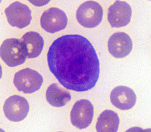
Lymphocyte
- The
appearance of lymphocytes in blood smears from guinea pigs is similar
to that in other species. Small, well differentiated lymphocytes
predominate and are slightly larger than RBCs. The larger lymphocytes
are almost twice as large and may have azurophilic granules, as in the
rabbit (Fig. 6).
|
|
|
Figure
6. Appearance of large and small
lymphocytes in a stained blood smear from a guinea pig. The large
lymphocyte at left has several metachromatic granules in the area of
nuclear indentation.
|
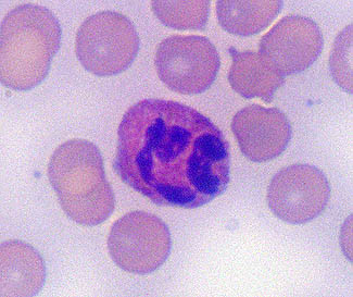
Heterophil
- Heterophils
of guinea pigs are 10 to 12 µm in diameter. The nucleus of individual
cells usually is purple, segmented, and has a dense chromatin pattern.
The nuclei of some heterophils in females have a "drumstick"
sex chromatin lobe. The cytoplasm has scattered acidophilic granules
that are smaller than those of eosinophils. More of the cytoplasm is
visible than in eosinophils.
|
|
|
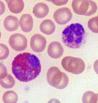
Figure
7. The heterophil (upper
right) has small, widely scattered, eosinophilic, cytoplasmic
granules compared to the eosinophil (lower left) that has numerous,
large, round, brightly eosinophilic, cytoplasmic granules.
|
Eosinophil
- Eosinophils
are slightly larger than heterophils. The nucleus is less segmented and
the cytoplasmic granules are larger, round, and bright red compared to
heterophils of this species. Granules usually completely fill the
cytoplasm (Fig. 7).
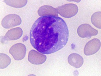
Monocyte
- Monocytes
are the largest leukocyte in circulation. These cells have a
variably-shaped nucleus, less condensed chromatin pattern, and
moderately abundant blue-gray cytoplasm. Compared to lymphocytes,
monocytes are larger and have darker, more abundant (Fig. 8).
|
|
|
Figure
8. Image of a monocyte to be
added
|
Basophil
- Basophils
are the same size as the other granulocytes. They have a purple, lobated
nucleus and variably-sized, purple granules in the cytoplasm.
|
|
|
Figure
9. Image of a basophil to be
added.
|
Foa-Kurloff
cells - Foa-Kurloff
cells are unique to guinea pigs and capabaras. These mononuclear cells
approximate the size of a large lymphocyte. They have a large, round,
purple nucleus that is often eccentrically located and blue cytoplasm.
The distinguishing characteristic of this cell is a very large,
slightly granular, magenta, cytoplasmic inclusion body (Fig. 10).
|
|
|
Figure
10. Foa-Kurloff cell in the
blood smear of a guinea pig. Notice the characteristic large,
slightly granular, magenta, cytoplasmic inclusion.
|
Comments
Concerning the Hematology of Guinea Pigs
The
predominant circulating leukocyte in healthy guinea pigs is the lymphocyte.
In contrast to the rabbit, basophils are rarely observed. Foa-Kurloff
cells may comprise 3 to 4% of leukocyte differential count.1
The published reference intervals for the total and differential
leukocyte counts are as follows:4 WBC = 8,220 - 14,000 cells
/µl; segmented heterophils = 1,350 - 3,650 cells /µl; band heterophils
= 0 – 10 cells /µl; lymphocytes
= 5,470 - 10,550 cells /µl; monocytes = 60-560 cells /µl; and basophils
= 0 – 20 cells /µl.
There
are few publications on leukogram changes of guinea pigs with naturally
occurring diseases. Guinea pigs experimentally infected with Trixacarus caviae
(guinea pig mange mites) developed a heterophilia, monocytosis,
eosinophilia, and basophilia.6 Although guinea pigs are
considered a tick-resistant species, they may develop an eosinophilia
and basophilia in response to infestation with Amblyomma americanum7
(lone star tick) and a significant basophilia in response to Rhipicephalus sanguineus (brown
dog tick) .8
Guinea pigs also have developed eosinophilia in response to Treponema pallidum
(syphilis) infections.9
References
1.
Moore DM: Hematology of Rabbits and Hematology of the Guinea Pig. In: Feldman BF,
Zinkl JG, Jain NC (eds): Schlam’s
Veterinary
Hematology, 5th ed, Lippincott Williams & Wilkins, 2000,
pp.1100-1110.
2.
Pouliot N, Maghni K, Blanchette F, et
al: Natural killer and lectin-dependent cytotoxic
activities of Kurloff cells: Target cell selectivity, conjugate
formation, and Ca++ dependency. Inflammation 20:647-671, 1996.
3.
Benson KG, Paul-Murphy J: Clinical pathology of the domestic rabbit:
Acquisition and interpretation of samples. Vet Clin N Am Exotic Anim
Pract 2:539-552, 1999.
4.
Campbell TW: Mammalian hematology: Laboratory animals and miscellaneous
species. In:
Thrall MA: Veterinary Hematology and Clinical Chemistry, 1st ed,
Lippincott Williams and Wilkins, 2004, pp. 211-224.
5.
Harcourt-Brown F. Textbook of Rabbit Medicine, 1st ed, Elsevier Science
Limited, 2002, pp.142-147.
6.
Rothwell TLW, Pope SE, Rajczyk ZK, Collins GH: Haematological and
pathological responses to experimental Trixacarus caviae infection in
guinea pigs. J Comp Pathol 104:179-185, 1991.
7.
Brown SJ, Askenase PW: Blood eosinophil and basophil responses in
guinea pigs parasitized by Amblyomma
americanum ticks. Am J Trop Med Hygiene 31:593-598, 1982.
8.
Szabo MPJ, Aoki VL, Sanches FPS, et
al: Antibody and blood leukocyte response in
Rhipicephalus sanguineus tick-infested dogs and guinea pigs. Vet
Parasitol 115:49-59, 2003.
9.
Wicher V, Scarozza AM, Ramsingh AI, et
al: Cytokine gene expression in skin of susceptible
guinea-pig infected with Treponema
pallidum. Immunology 95:242-247, 1998.
Acknowledgment
"Ranch
Rabbit", an acrylic painting by Malcolm Furlow,
is from the Creative Expressions Gallery
website and permission to use has been requested.
Web Design by Lois Klesa Morrison
|