![]()
Flatworms: rabbit as an intermediate
host
Esther van Praag, Ph.D.
|
|
MediRabbit.com is
funded solely by the generosity of donors. Every
donation, no matter what the size, is appreciated and will aid in the
continuing research of medical care and health of rabbits. Thank you |

Warning: this file contains pictures that may be
distressing for some persons.
Taenia pisiformis
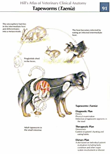
Tapeworm Taenia pisiformis
is a common parasite of carnivores such as dogs, foxes and, on occasion,
cats. It is found all over the world, predominantly in rural regions. The development of the parasite is a
two-stage process: 1. An adult stage, as an adult parasite in
the definitive host, the dog. The parasite lives in the small intestine (duodenum
jejunum and ileum) of the
dog and may reach a length of 2 meters (approximately 79 inches). Mature
segments of the tapeworm (protoglottid) containing
mature eggs are shed along with the feces. 2. An intermediate stage in an herbivorous
host where the larval stage is found (mesacestoide). Rabbit or other
lagomorph species may ingest the eggs while grazing contaminated grass.
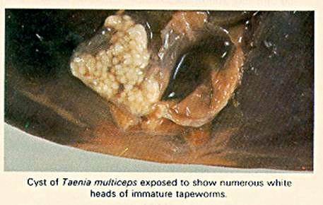
www.powhatananimalhospital.com/disease/tapeworms2.jpg The intermediate stage (mesacestoide) is marked by the presence of a bladder-like
structure in the abdominal/peritoneal cavity and liver. This intermediate
larval stage is known as Cysticercus pisiformis. The development of the larva is blocked,
and they will survive in the bladder structure. Tapeworm-related cysts are
viable and can reach a size of up to 2-3 cm in diameter, with rare cases
reaching 8 cm or larger. The progression to the adult stage is only possible following the ingestion of an infested rabbit's viscera by a dog, fox, or cat. The development of the larva will continue until it reaches adulthood.
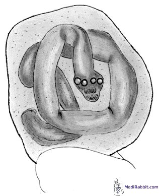
Richard Hoop Blister-like
mature cysticerci (green arrows)containing a larva of the tapeworm Taenia
pisiformis in the body cavity of a rabbit.
Detail
of a cyst, showing the larval taperworm Cysticercus pisiformis surrounded by fluids. The parasitic larvae use the hepatic
portal vein to invade the liver of rabbits. The migration phase of the larva
is usually accompanied by focal granulomatous hepatitis-like symptoms, which
include inflammation of the liver, local hepatocellular necrosis, and hepatic
scarring if the condition becomes chronic. A thorough microscopic analysis of
the tissue reveals the presence of necrotic tissue, blood, degenerative
granulocytes, giant cells, and mononuclear cells. Fibroblastic transformation
of the tissue may be present, and the affected tissue will be replaced by
connective tissue. After 15 to 30 days, the larva will migrate to the hepatic
parenchyma and form cysts. A severe infestation can result in
chronic extreme weakness or sudden death.
There is a possibility of aberrant
migration. Some of these cysts have been found in the peritoneal fluids or in
the lungs of a rabbit, filling almost all the cavity of the lung and causing
respiratory distress. Cysticercosis of the brain can cause seizures, increased intracranial pressure, and altered mental
status. The X-rays indicate the presence of hydrocephalus, aseptic
meningitis, and/or calcified cysts. CT scanning and MRI are the preferred
diagnostic tools for definitively identifying cysts, often accompanied by
ring-enhancing lesions or hydrocephalus. Given the fact that MRI cannot
clearly show the calcified pocket, the use of a contrasting dye administered
intravenously is recommended to better visualize the inflamed and destroyed
regions. As long
as there is no direct contact or ingestion
of the cysts containing the larva (unlikely in pet rabbits), contamination is
not a concern. Treatment can be attempted with
praziquantel. Multiceps serialis
This parasitic worm is also referred to
as Taenia serialis. As with Taenia pisiformis,
the development of the parasite follows a two-stage pattern: an intermediate
stage in hares or wild rabbits, and an adult form in dogs and cats. The
occurrence of this condition is uncommon among domestic rabbits. In rabbits,
cysts of Multiceps serialis are found
in the subcutaneous tissues and muscle mass. The cysts are characterized by
their elongated form, and the larva is clearly visible.
www.unbc.ca/nlui/wildlife_diseases/taenia_multiceps.htm
The development stage is blocked at the
cyst stage in rabbits. Maturation and development into the adult stage is
only achieved through the consumption of an infested rabbit. There is a potential risk of human
contamination, which can occur when there is direct contact with the cysts or
ingestion of the intermediate larva (e.g., from hunted hares or rabbits, or
contact with the digestive tract). The
risk of contamination is minimal when it comes to pet rabbits. Acknowledgement
I would like to express my gratitude to Professor Richard Hoop (Institut für Veterinärbakteriologie,
University of Zurich) for his permission to use his picture related to Taenia
cysts in rabbits Further information:
Manning
et al. The biology of the laboratory rabbit. 2nd ed. London, UK,
1994. Maynard
A. Novlesky MA, Dyer WG. Helminths of the Eastern Cottontail
Rabbit, Sylvilagus
floridanus, from North Dakota, by © 1970 The University of Notre
Dame. Pinto
RM, et al. Helminths of rabbits (Lagomorpha, Leporidae) deposited in the
Helminthological Collection of the Oswaldo Cruz Institute. Rev. Bras. Zool. 2004, v. 21, n. 3, pp. 599-604. Soltysiak
Z, Bednarski M, Piekarska J. Wagrzyca watroby królika. Medycyna Wet. 2007,
63:1255-1257. |


e-mail: info@medirabbit.com
