![]()
Passalurus ambiguus
Esther van
Praag, Ph.D.
|
|
MediRabbit.com is
funded solely by the generosity of donors. All
donations are greatly appreciated and will contribute to ongoing research on
medical care and rabbit health. Thank you |

|
This
parasite is also known under the name Oxyuris ambigua. This parasite
is a common cosmopolitan pest that has been found to infest wild and pet
rabbits, cottontails, and hares. In the USA, the presence of P.
nonanulatus has also been observed in rabbits on occasion. Auto-infection
is a common occurrence, typically resulting from the ingestion of the eggs
with food. Passalurus sp. has been observed to inhabit the mucosa of
the small intestine and the cecum during its juvenile stages. As it matures,
the adult worms move to the anterior part of the cecum and the large
intestine of rabbits. Passalurus
ambiguus is specific to lagomorphs and does not
represent a public health risk. The life cycle of Passalurus sp.
is direct: the eggs are ingested by the animal. Eggs typically have a flat
side and measure about 100*43 micrometers. During their development,
resistance against dehydration increases. The larvae will hatch from the eggs
and develop in the mucous layer of the small intestine and the cecum, where
they will develop into mature adults. Two molts have been documented: the
first occurring after 24 hours and the second on the third day.
Adult worms exhibit size variations, with
males measuring approximately 5 millimeters and females measuring
approximately 10 millimeters. The females, distinguished by a long and narrow
tale, are marked with approximately 40 circular, cuticular striations. It
seems that the female worms deposit the eggs around the anus. The lifespan of
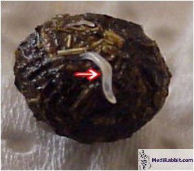
the worms is approximately 106 days. Clinical signs
Passalurus sp. parasites are non-pathogenic
organisms, and their presence is usually asymptomatic, even in severe
infestations. A rabbit may be infested with over 1,000 parasitic worms. Eggs
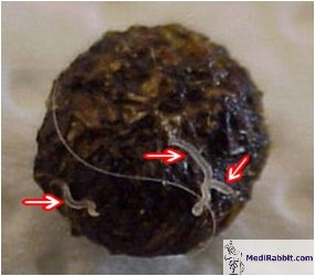
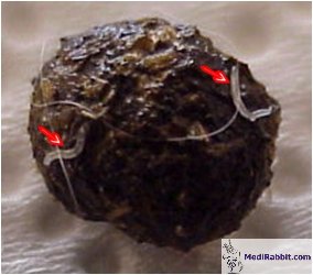
and, on occasion, live adult worms can be identified in freshly excreted
feces. Once out of the rabbit body, the worms will dry quickly and will not
be visible after five minutes. It has
been observed that the female worms that emerge from the anus of rabbits
contain eggs in the gastrula stage that can develop into an infective stage
within the environment. The
presence of mucus threads between the droppings can be a serious indication
that a rabbit may be infested by intestinal worms. It is important to note
that this condition should be differentiated from mucoid enteritis. An
overpopulation of the nematode worms in the digestive tract can lead to
stasis and cecal impaction, as well as pain and gas production. If not
treated, the stasis can become chronic, with symptoms recurring every four to
six weeks. During the necropsy, Passalurus
sp. worms were identified within the lumen of the cecum, as well as in the
crypts and mucosa of the colon. The site where the worms were located
exhibited inflammatory changes and dystrophic modifications. The most
significant inflammatory and dystrophic changes were observed in the cecum.
Furthermore, signs of vascular dystrophy were observed in the hepatic and
renal parenchyma.
Diagnosis
The
presence of intestinal parasites is determined by a fecal flotation test. In
rare cases, the fecal flotation test result of heavily infested rabbits can
return negative. If left untreated, the presence of worms can often be
observed in the droppings. Treatment
Further Information
H. Boecker (1953) Die Entwicklung des
Kaninchen Oxyuren Passalurus ambiguus.
Zeitschrift für Parasitenkunde 15: 491-518. S. Brown (1993) Rabbit Drug Dosages. Rabbit
Health News 10: 6-7 J. Burke (1994) Clinical Care and
Medecine of Pet Rabbits. In: Proceedings of the Michigan Veterinary
Conference, pp 49-77. D. Duwel and K. Brech (1981)
Control of Oxyuriasis in Rabbits by Fenbendazole. Lab. Anim. Sci. 15:
101-105. A.B. Erickson (1944) Helminth
Infection in Relation to Population Fluctuations in Snowshoe Hares. J. Wildl.
Manage. 8: 134-153. E.V. Hillyer, K.E. Quesenberry
(1997) Ferrets, Rabbits, and Rodents. Clinical Medicine and Surgery. W.B.
Saunders Company pp. J.P. Hugot (1984) L’Insémination
Traumatique chez les Oxyures de Dermpotères et de Léporidés. Etude
Morphologique Comparée. Ann. de Parasitologie Humaine et Comparée 59:
379-385. M.A. Palimpsestov, R.S. Chebotarev
(1935) Zur Frage des Therapie bei Passalurose (Passalarus ambiguus) des Kaninchen. Tierärtzliche Rundschau 41:
709-711. N.M. Patton, K.W. Hagen, J.R. Gorham,
R.E Flatt (1986) “Domestic Rabbits: Diseases and Parasites. “Pacific
Northwest Extension Publ. Oregon, Idaho, and Washington. K.I. Skrjabin, N.P. Shikhobalova, E.A. Lagodovskaya (1960)
Oxyurata of Animals and Man. Part I. Oxyuroidea. In: Skrjabin,
K.I. (ed.), Essentials of Nematology, The Academy of Sciences of the
U.S.S.R., Moscow (published by the Israel Program for Scientist Translations,
Jerusalem, 1974). J. Theodoris
(1979) Contribution to the study of parasites in the rabbits of Northern
Greece. hellinike Kteniatrike (Hellenii Veterinary Medecine) 22: 181-183. T.L.H Tsui and N.M. Patton (1991)
Comparative Efficiency of Subcutaneous Injection Doses of Invermectin against
Passalurus ambiguous in rabbits J.
Appl. Rabbit Res. 14: 266-269. N.B. Walden (1999) Rabbits: a
Compendium (The T.G. Hungerford VADE MECUM series for Domestic Animals:
Series C.13). Post Graduate Foundation in Veterinary Science, University of
Sydney, Sydney. J.P. Wiggins, M. Cosgrove, H.
Rothenbacher (1980) Gastrointestinal Parasites of Eastern Cottontail Rabbits
(Sylvilagus floridanus) in Central Pennssylvania. J. Wildl. Dis.
16:541-544. Fujiwara H, Uchida K, Takahashi M.
[Occurrence of granulomatous appendicitis in rabbits] Jikken
Dobutsu. 1987; 36(3):277-80. Shirokova EP, Grishina EA. [Microstructural changes
in the organs of the rabbit with passaluriasis] Med Parazitol (Mosk) 1997;
(2):18-21. |
||||||||||||||||||||||

e-mail: info@medirabbit.com
