![]()
Nematodirus leporis
Esther van Praag,
Ph.D.
|
|
MediRabbit.com is funded solely by the generosity of
donors. Every donation, no matter what the
size, is appreciated and will aid in the continuing research of medical care
and health of rabbits.
Thank
you |

|
The
parasitic nematode Nematodirus leporis generally experiences a peak infestation
during the spring months. It is a thin-necked intestinal worm that is
occasionally found in wild rabbits and hares. Pet rabbits residing in
temperate, cold, or elevated environments are also susceptible to infestation.
Additionally, the presence of N. neomexicanus,
N. arizonensis, and N. triangularis
has been documented in wild rabbits. There is no reported public health risk
related to this parasite The life cycle of Nematodirus
leporis is direct, with no intermediate host.
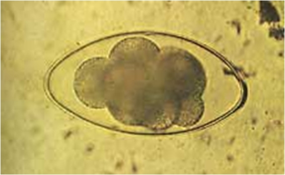
The eggs, which are thick-layered, are significantly larger (250*100 micrometer)
compared to those from other Trichostrongylidae
species. They demonstrate exceptional resistance to desiccation, freezing
conditions, and snow. Typically, the eggs have begun to divide rapidly, with
one to eight dark cells being visible under a microscope. The development of
the larvae is generally slow, with a duration of up to two months in
temperate climates. It is contingent upon the environmental humidity and
temperature. During the hatching process, the larva sheds the first-stage
cuticle, which is left behind in the eggshell. The L3 larva remains within
the eggshell, which provides protection against adverse environmental
conditions. This allows the L3 larvae to survive for up to one year in
pasture fields. After the L3 larva is ingested by the host, it will exsheath and move to the paramucosal
lumen of the small intestine. There, it will molt into the L4 and immature
adults.
The adult parasite is slender, measuring
30 mm in length. Its body shape is curled and presents 18 longitudinal
striations. The anterior part is reduced, with an inflated cuticle that is
typically striated. The anterior part is inflated with a noticeable dorsal
esophageal spicule. The male worms possess a bursa with two large lateral
lobes, covered with mediolateral and caudolateral striations. The female's
tale ends abruptly.
Clinical signs
The
clinical and pathological signs of Nematodirus
sp. infestation are only noticeable in severe cases, leading to diarrhea,
weight loss, and performance issues. The necropsy revealed that the high
number of worms form clumps that resemble cotton wool. These clumps are
usually intertwined around the intestinal villi, which causes atrophy,
degeneration, and necrosis of the surface enterocytes. The
presence of a Nematodirus sp. infestation
can be determined through fecal flotation to identify the significant
presence of Strongyle-type eggs and adult worms. Treatment
Further
Information
Audebert F, Cassone J, Kerboeuf D,
Durette-Desset MC. The life cycle of Nematodiroides zembrae
(Nematoda, Trichostrongylina) in the rabbit. J Parasitol. 2002; 88(5):898-904. Hoste H, Mallet S, Fort G.
Histopathology of the small intestinal mucosa in Nematodirus spathiger
infection in rabbits. J Helminthol. 1993; 67(2):139-44. Hoste H, Fort G. Experimental
infections with Nematodirus spathiger in rabbits. J Helminthol. 1992;
66(3):227-30. Andrews CL, Davidson WR.
Endoparasites of selected populations of cottontail rabbits (Sylvilagus
floridanus) in the southeastern United States. J Wildl Dis. 1980;
16(3):395-401. Knight RA. Effect of dexamethasone
on experimental infections of Trichostrongylus affinis and Nematodirus
spathiger in rabbits. J Parasitol. 1977; 63(5):957-8. Jansen J. Where does Nematodirus
battus Crofton & Thomas, 1951, come from? Vet Rec. 1973;
92(26):697-8. Gallie GJ. The pathogenicity of Nematodirus
battus in weaned and unweaned laboratory rabbits. J Helminthol. 1973;
47(4):377-88. Gallie GJ. The development of
acquired resistance and age resistance to Nematodirus battus in the
laboratory rabbit. J Helminthol. 1973; 47(4):369-76. Mapes CJ. Bile and bile salts and
exsheathment of the intestinal nematodes Trichostrongylus colubriformis
and Nematodirus battus. Int J Parasitol. 1972; 2(4):433-8. Gallie GJ. Development of the parasitic stages of Nematodirus
battus in the laboratory rabbit. Parasitology. 1972; 64(2):293-304. |
|||||||||||


e-mail: info@medirabbit.com
