![]()
Graphidium
strigosum
Esther van
Praag, Ph.D.
|
|
MediRabbit.com is
funded solely by the generosity of donors. Every
donation, no matter what the size, is appreciated and will aid in the
continuing research of medical care and health of rabbits. Thank you |

|
This cosmopolitan parasite is
primarily observed in the wild rabbit (Oryctolagus cuniculus) and in
the Leporidae family, which includes the hare (Lepus europaeus, Lepus
capensis). Rabbits are likely the primary host species, as they exhibit a
higher tolerance for Graphidium strigosum compared to hares, which
often develop significant stomach lesions. It is therefore hypothesized that
the presence of this worm in hare populations is contingent on the presence
of a population of wild rabbits. A study has revealed a correlation between Graphidium
strigosum and Trichostrongylus retortaeformis in wild rabbits.
House rabbits are susceptible to infestation by green forage, which is
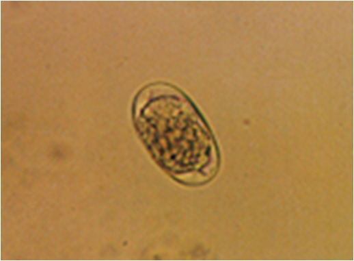
contaminated with eggs and infectious larvae. Current knowledge of the biology and life cycle of this parasite
is limited. The eggs measure approximately 95*50 micrometre in size. They are
laid in the morula stage. In optimal environmental conditions, the larvae
will hatch approximately 10 hours later. The L2 stage is typically reached
within 2 to 3 days. Ensheathed L3 larvae are capable of infecting hosts and
migrate along the herbage according to the time of day. Specifically, they
move to the tip at dusk and downwards when exposed to sunlight and heat. This
behaviour continues until they are ingested by their host. Male and female
adult worms are red with many longitudinal lines and transversal striations.
The males measure approximately 12 millimetres in length, while the females
average 16 millimetres. Males are characterized by the possession of paired,
slender spicules and a well-developed copulatory bursa.
Clinical signs
The clinical signs are
comparable to those observed in cases of gastritis. Signs of a severe
infestation include catarrhal gastritis with fibrosis and extreme
inflammation of various parts of the intestinal tract (stomach, small
intestine, cecum). Necropsy findings indicate that
L4 stage worms are coiled within the ducts of the gastric glands in the
fundus region. Adult worms are usually found in the mucus layers. It is
important to note that their heads are generally buried in the stomach
grooves, without attachment to the mucosa. Treatment
Further
Information
B. Boag (1987) The Helminth Parasites of the Wild Rabbit Oryctolagus cuniculus and the brown
hare Lepus capensis from the Island
of Coll, Scotland. J. Zool. 212: 352-355. B. Boag and H.H. Kolb (1989) Influence of the Host Age and Sex on
Nematode Populations in the Wild Rabbit (Oryctolagus
cuniculus L.). Proc. Helminth. Soc. Washington 56: 116-119. Brookhuizen and Kemmers (1976) The Stomach Worm Graphidium strigosum (Dujardin)
Railliet and Henry, in the European Hare, Lepus
europaeus Pallas. In: Pielowski, Z. and Pucek Z. (eds) Ecology and
Management of the European Hare Populations. Panstwowe Wydawnictwo Rolnicze i
Lesne, Warshaw, Poland, pp 157-171. J.D. Dunsmore, M.L Dudzinski (1968) Relationship of Numbers of
Nematode Parasites in Wild Rabbits, Oryctolagus
cuniculus (L.), to Host Sex, Age and Season. J. Parasitol. 54: 462-474. E.A. Nickel and W. Haupt (1986) Experimental Studies on the
Course and Consequences of Infection with Graphidium
strigosum (Nematoda, Trichostrongylidae) in Oryctolagus cuniculus. Agnew. Parasitol. 27, 215-219. E.J.L. Soulsby (1968) “Helminths, Arthropods, and Prorozoa
of Domestic Animals”. Williams and Wilkins, Baltimore, Maryland. R. Wetzel, K Enigk (1937) Zur Biologie von Graphidium strigosum, dem Magenwurm
der Hasen und Kaninchen. Deutsch. Tierärtzliche Wochenschrift 45: 401-405. |
e-mail: info@medirabbit.com
