![]()
Be careful, rabbit
pseudotuberculosis
is
transferable disease that can be passed on to humans
Michel Gruaz
(Article published in the animal
journal Tierwelt and graciously allowed to share
here in MediRabbit.com by M. Gruaz)
|
|
MediRabbit.com is funded solely by the generosity of
donors. Every donation, no matter what the size,
is appreciated and will aid in the continuing research of medical care and
health of rabbits.
Thank you |

Warning: this page
contains pictures that may be distressing for some persons.
This sole and unique reason justifies
informing rabbit owners about this disease, which is rare but is observed
from time to time. It is important to acknowledge this, as it is somewhat out
of the ordinary.
Over the course of
several years, I had three cases within a few months of each other.
Consequently, the author of this article is able to report on the clinical
signs of the disease. This condition primarily affects adult animals or those
that are several years old. The rabbit's appetite diminishes progressively,
and after a few days, it will only consume small amounts of fresh food before
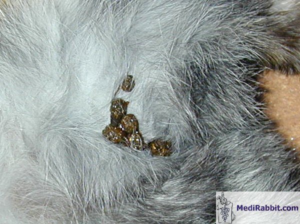
completely ceasing to eat. The droppings become progressively smaller,
harder, and stickier. The ventral abdomen is soft to the touch, and during
palpation, it is possible to feel that the contents of the abdominal cavity
decrease.
The animal rapidly
loses weight. If the animal is not euthanized, death will occur within a few
days. Following the presentation of the post-mortem images to one
veterinarian, the images were subsequently reviewed by Dr. med. vet. Richard
K. Hoop of the Institute of Veterinary Bacteriology at the University of
Zurich (Switzerland) diagnosed the condition as pseudotuberculosis. These
experts advised the author of the potential risks of transference to humans
and recommended against touching one's mouth after handling a sick rabbit. It
is important to note that no additional cases have been observed among the
rabbit population under the author's care.
According to Boucher et Nouaille the disease is rare
in rabbits
Yersiniosis or pseudotuberculosis is a rare disease in rabbit
husbandry. However, it should be noted that it is capable of infecting birds,
such as the common wood pigeon, as well as rodents, including guinea pigs.
There is a risk of infection when there is contact between the latter and
rabbits. The disease has been observed in farm rabbits living in hutches and
in pet rabbits living in the vicinity of birds infected by the bacterium.
Wild rabbits and hares can be a source of infection. Hares are highly susceptible
to yersiniosis. In this species, the disease is frequently observed. It is
the primary cause of mortality in certain regions of France and Germany. The
disease is considered zoonotic, meaning it can be transmitted to humans. This
disease primarily affects boys between the ages of 8 and 13. The symptoms are
comparable to those associated with appendicitis. These symptoms may be
accompanied by skin inflammation and redness. It is important to note that
both humans and animals can be affected by either a generalised
infection and severe septicaemia, or by localised infections in the lungs or eyes. The septicemic
form is fatal within 24 to 48 hours, while the chronic form may take between
two and three months to become fatal, although many survivors have been
documented.
A bacterium called Yersinia
is causing the disease
This
infection is caused by Yersinia pseudotuberculosis, a bacterium that was
discovered in 1883 on a guinea pig inoculated with the nodule of a child who
died of tuberculous meningitis. It is a small, cosmopolitan, Gram-negative
bacterium that is rod-shaped. This bacillus or bacterium is present in
infected and healthy animal and human individuals. It is primarily birds and
rodents that act as the reservoir for this disease. However, it is important
to note that this bacterium can also be found in various environmental
sources, including soil, water, and food contaminated with the excreta of
infected animals. In rabbits, the bacterium has been observed in fecal
samples as well as in cecal droppings. It has the capacity to survive in the
soil for over a year. It is capable of multiplication at temperatures ranging
from 4 to 10°C. This ability may be linked to the rise in the number of
infected rabbits during the winter season. The soil is the primary source of
contamination. It is therefore imperative that particular care is exercised
when burying a dead rabbit. Caution is needed when applying manure to the
soil. It is imperative to ensure that bodies are not decaying in a pile of
manure. It is possible for this bacterium to spread back into the soil in
this manner.
Following ingestion, the bacterium reaches the intestine, where it
begins to multiply. At 37°C, there will be a rapid increase in the bacterial
population, with bacteria beginning to invade the lymphatic nodules
associated with this organ. In the septicemic form of the disease, the rabbit
will die rapidly and without noticeable clinical signs. In the non-septicemic
forms, the rabbit will exhibit signs of fatigue, including emaciation and
exhaustion. It is possible that the subject will also experience diarrhea.
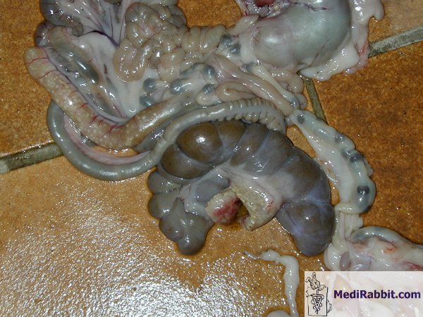
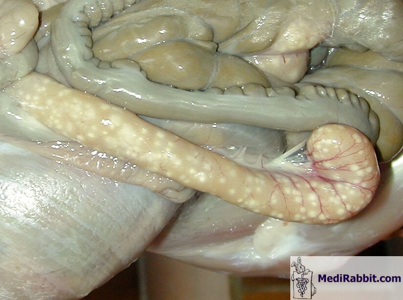
During palpation, the nodules exhibit signs of hypertrophy. The liver
displays a pattern of fairly hard nodules. The intestine may exhibit necrotic
regions. In some cases, the kidneys and lungs are affected. It should be
noted that not all animals die.
Suspicion, onset and passing over of
the disease
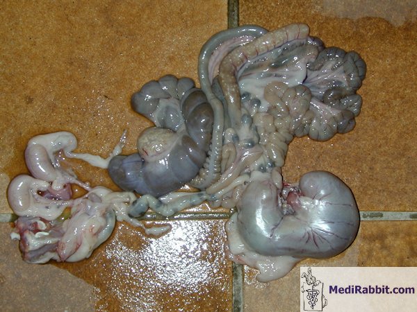
Necropsy results may provide useful diagnostic insights. The results
of this examination indicate the presence of hypertrophy of the lymphatic
nodules and a spleen that is twice to three times larger than normal. The
kidneys, spleen and intestines display minor white/yellowish nodules on their
surface. These nodules bear a resemblance to those observed in cases of
tuberculosis, hence the term "pseudotuberculosis". A
bacteriological examination of the affected organs (kidney, spleen, liver,
intestine, bone marrow) is the sole diagnostic tool that can provide a
conclusive diagnosis of the disease. It is imperative for rabbit owners who
also have birds, hamsters, guinea pigs, or poultry in the same barn to
educate themselves on fundamental hygiene practices. The feeding of grains
left over by birds living in hutches to poultry is a well-known source of
contamination by Yersinia bacteria in the barnyard. It is also important to
look for possible contamination of the soil and plants. Contamination is
often of an oral and digestive nature.
The incubation period is long, with an average duration of 15 days.
The bacteria then reach the lymphatic system, and nodules appear on different
organs. Following the ganglionic stage, the liver is invaded and becomes the
starting point for septicemia.
Prevention is always more desirable
than treatment
It is recommended that hutches, pens, and the living environment of
the rabbit be disinfected, as well as the soil. Walls can be thoroughly
cleaned. It is imperative that the water is treated with an antibacterial
product in order to eradicate the bacterium before disposal. Accessories and
fodder must be destroyed. These objects can be soaked in a bactericidal
solution, which is widely available in drugstores, for a minimum of one hour.
The drinking water of the rabbits is treated with a disinfectant that is
based on chlorine. It is imperative to address the issue of rodents in a
coordinated manner. It is also imperative to meticulously inspect other
breeding sites. It is recommended that prophylactic measures be taken. The
treatment itself involves the administration of enrofloxacin (Baytril), at a dosage of 15 mg/kg of fresh weight, for a
period of 8 days. Following a 10-day cessation of treatment, a second
antibiotic regimen is administered for an additional 8 days. A
bacteriological study, also known as an antibiogram, is a methodical analysis
that evaluates the sensitivity of bacteria to various antibiotics. This test
is essential for determining the efficacy of other molecules.
More Information
Maladies des
lapins de Boucher et Nouaille
Textbook of Rabbit Medicine, Frances
Harcourt-Brown, Oxford,
The Biology of the Laboratory Rabbit, Patrick J.
Manning, Daniel H. Ringler and Christian E. Newcomer,
Vetstream,
https://www.vetstream.com/lapis/Content/Freeform/fre00324.asp
Zoonoses and Communicable Diseases Common to Man
and Animals: Bacterioses and Mycoses v. 1 - PAHO
Scientific Publications S. No. 580, Pedro N. Acha,
Boris Szyfres.
|
|||||||||

e-mail: info@medirabbit.com
