![]()
Late opening of eyelids in newborn rabbits
Consequences
Esther van Praag, Ph.D.
|
MediRabbit.com is funded solely by the
generosity of donors.
Every donation, no matter what
the size, is appreciated and will aid in the continuing research of medical care
and health of rabbits.
Thank you
|

|
Newborn rabbits are nidicole and, as a consequence, born
immature, naked, deaf and blind. Their eyelids are closed. It is only after
their opening, around the age of 10 days, that the consequences of a late opening
or deformations of the eyelids become visible.
Rabbits are born
with the lower and upper eyelids stuck shut to protect the eye - an entity that includes
the optic nerve and the eye globe. Indeed, the development of the eye is
not complete in newborn rabbits; it is in no way a miniature version of the
adult vision organ. The size, shape, but also the proportions of the
different parts of the eye change during the first weeks of life of the
newborn rabbit. The proportions between the anterior chamber and the
posterior chamber of the eye are very different from those of the
adult. The surface of the retina - the organ that is sensitive to vision at
the back of the eye, increases and becomes more and more innervated. The
cornea and the conjunctiva, respectively the transparent layer covering the
eyeball and the transparent mucous membrane lining the inside of the eyelids,
are also immature and will continue their development after the birth of the
rabbit.
MediRabbit Anatomy of the eye of a healthy rabbit Opening of the eyelids The opening of the eyelids happens usually on the 10th
day after birth. A small split appears in the inner corner of the eye that progresses slowly from the outer
cutaneous portion of the eyelid to the conjunctival inner surface. Ligaments and muscles of the eyelids also participate in the appearance of the
split. Once the eyelids are separated, the free edges gradually harden. Eyelashes
grow. The sebaceous glands located on the inner edge of the eyelids become
functional and release secretions to lubricate the eyelids. The full opening
of the split may take a few days. When completed, the upper and lower eyelids
protect the cornea against foreign objects, trauma, light or wind. They help maintain
a proper hydration of the cornea by distributing the tear fluid - secreted by
the various lacrimal glands via the lacrimal duct, over its surface. The opening of the tear
duct is visible inside the lower eyelid, when it is delicately pulled
forward.
Rabbits have a third eyelid or nictitating membrane. Its
active or passive movements protect the cornea and the conjunctiva. The
nictitating membrane has an accessory lacrimal gland which
contributes to the aqueous portion of the tear film. Rabbits beat their
eyelids only 10 to 12 times per hour.
Cornea and lens The organization of
corneal tissue begins during the development of the embryo, with a very precise organization
of collagen fibrils. This phase is critical as it ensures the transparency of the
cornea in adult rabbits. At birth, the cornea is opaque. Its development is
linked to the opening of the eyelids. At this time, the state of hydration
and vascularization differs from that of adult rabbit and important changes
will take place. The thickness of the cornea will decrease after the
opening of eyelids. This process continues for a few weeks, then the
membrane thickens and becomes similar to that observed in adults. These
changes are accompanied by a decrease in the density of cells in the cornea. The
lens of the eye also changes. During the first two weeks of life of the
rabbit, the lens shows a spectacular growth and reaches 50% of the size of
the adult rabbit. During this period, the lens accumulates cholesterol
molecules and gradually becomes transparent. In young rabbits with a late
opening of the eyelids, irreversible opacity of the cornea and/or lens is
often observed. It is assumed that a delay of eyelid opening disorganizes the development
of these tissues and disturbs their transformation, which leads to an opacity of the cornea and/or the lens. Very little information is
available on this topic.
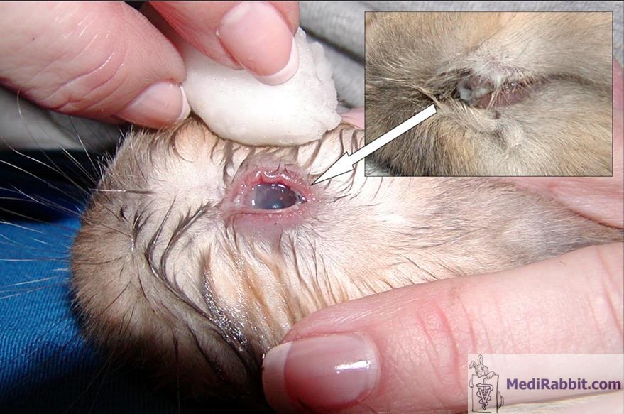
C. Wayne Wright Closed eyelid et secretion of a thick
fluid is often a sign of conjunctivitis Late opening of the eyelids and possible infection The eyes of the newborn
rabbits are very sensitive organs and the risk of bacterial infection is high
during the first weeks of life. If the opening of the eyelids is late, beyond the 10th day of life, there is a high risk of developing a neonatal infection
of the conjunctiva and the outer layer of the cornea . Any suspicious
inflammation, protuberance or presence of pus must be taken seriously. Bacteria
such as Staphylococcus spp. or Streptococcus spp. are often
responsible for these infections. The newborn comes into contact with these
pathogens during parturition, when passing through the urogenital system of the
doe, but also in the nest, especially in the colder season. Characteristic signs of such infections are eyelids pointing outwards
because of inflammation or accumulation of pus in the inner eye corner, and the
presence of crusts or thick, yellowish-colored secretions between the
eyelids. The conjunctiva may become inflamed, reddish and the cornea is
opaque. In severe cases, the structures of the eye may be affected. A
superficial lesion of the cornea may develop into a painful ulcer; it may be
accompanied by opacification of the surface of the cornea and lens. This
opacity is often temporary and regresses slowly with appropriate treatment.
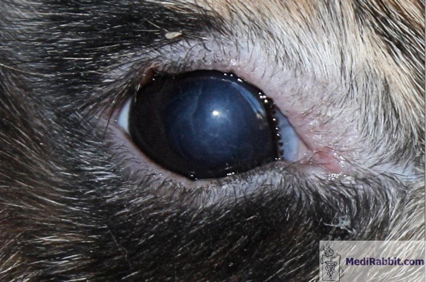
Michel Gruaz Opaque cornea after a late
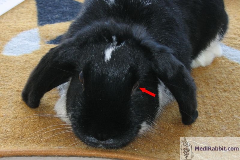
opening of the eye in a 3 weeks old rabbit A retraction of the
eyeball inside the eye-socket is sometimes observed after a late opening of
the eyelids.
Michel Gruaz Retracted eye in a
lop rabbit Treatment of non-opened eyelids
If a treatment is started quickly, the chances of total
healing are good. When the eyelids are not showing any sign of opening (split near in the inner corner), or
remain closed beyond the 10th day, it is necessary to open them manually.
The eyelids are moistened with sterile saline solution and separated.
Very gently to avoid damaging or tearing the eyelids. Once open, the eyelids and the eye are washed with a sterile saline solution
to eliminate any cellular waste or foreign body. In case of infection, it is
necessary to remove the infectious waste material at regular intervals and to
administrate antibiotic eye drops. Warm compresses placed on the eyelids will prevent
them from closing together again. It is important to keep the cornea moist,
especially if the infection prevents the twitching of eyelids. This can be
ensured by the administration of "artificial tears" drops
for human use that do not contain preservatives. If the surface of the cornea
is damaged, scarring may lead to the formation of a whitish veil. Even if it
is unsightly, the adult rabbit suffers at most from a small non-painful
visual discomfort.
It is important to
examine the other members of a litter as a newborn rabbit is rarely affected
alone.
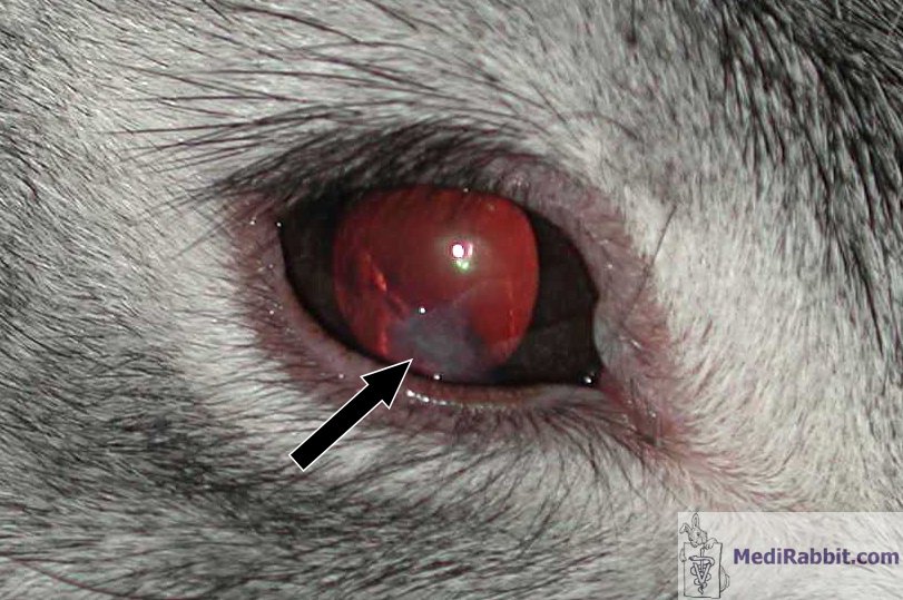
MediRabbit Scar on the cornea of an adult rabbit,
after a traumatic lesion of the cornea when she was a few weeks old Acknowledgements Huge thanks to Michel Gruaz (Suisse) and C. Wayne Wright
(USA) for their pictures and their permission to use them in MediRabbit. |

