![]()
Aberrant
corneal occlusion or pseudopterygium
Esther van Praag, Ph.D.
|
MediRabbit.com is funded solely by the
generosity of donors.
Every donation, no matter what
the size, is appreciated and will aid in the continuing research of medical care
and health of rabbits.
Thank you
|

Warning: this file
contains pictures that may be distressing for people.
A variety of names - pseudopterygium, precorneal membranous
occlusion, aberrant conjunctival overgrowth, or conjunctival
centripetalization - have been given to the aberrant growth of conjunctival
membrane tissue. The condition is progressive and extends from the bulbar
conjunctiva onto the cornea of the rabbit eye. The condition is very poorly
described in veterinary literature.
The etiology of the disease is unknown. It appears the result of an
inflammatory process that leads to the adherence of a fold of the conjunctiva
to the cornea, near the border of the cornea and the sclera (corneal limbus)
or more centrally. Breed, age, and sex of the rabbits seem to play a role, as
male dwarfs, aged between 5 and 12 months are more particularly affected. It
appears congenital in some cases. A further possible cause for
pseudopterygium may be ultraviolet radiation.
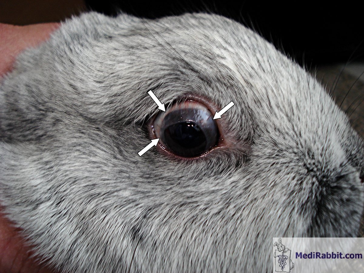
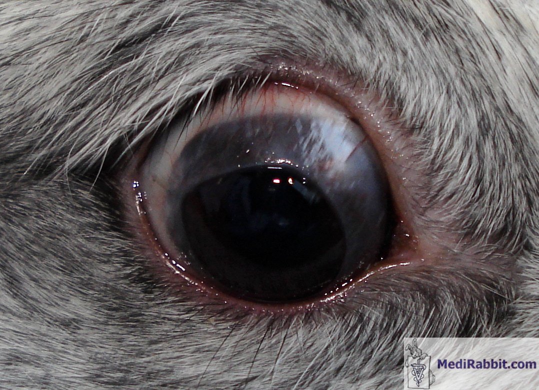
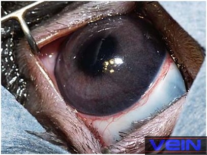
Stefan Röthlisberger
The conjunctival membrane covers partially the cornea. Vascularisation of the membrane is well visible
Clinical signs
"Pseudo" in the term pseudopterygium refers to the fact
that the conjunctival membrane is not adhering to the underlying cornea, but
growing over it. In rare cases, the membrane may be loosely attached to the
cornea, but can be easily separated without causing damage. It can cover only
a small part of the cornea with an annular peripheral opacification of the
cornea, or cover it almost fully, leading to blindness. When the membrane is
sectioned from the outer edge to the corneal limbus, it usually retracts back
to its normal position. The condition can be unilateral or affect both eyes.
Pseudopetrygium can be accompanied by pain if the cornea is damaged
by, e.g., a foreign body or a piece of hay stuck under the membrane.
Two cases of pseudopterygium in rabbits have been described by
Arnbjer. In the first case, the conjunctival membrane was detached with a
blunt instrument from the underlying cornea, followed by injection of
methylprednisolone acetate in the subconjunctival membrane. The eye was
treated with steroid/antibiotic eye drops during 3 week. In the second case,
the only treatment was topical antibiotics after the detachment of the
membrane; it grew back within weeks.
Treatment
As surgical removal of the overgrown conjunctiva leads to recurrence,
the condition can be left untreated if it does not hinder the sight of the
rabbit, and does not cause pain.
The surgical procedure that leads to the best results includes
suturing the fold back to the eyelid, to the sclera, or to the loose arching
folds connecting the conjunctival membrane lining the inside of the eyelid
with the conjunctival membrane covering the eyeball (fornix conjunctivae)
on a fully anesthesized rabbit. This can be done with resorbable Dexon or
Vicryl sutures 5.0 or 6.0. Post-surgical care includes the daily
administration of topical cyclosporine 0.2% and corticosteroids (e.g.
dexamethasone 0.1%) during a few weeks.
Rarely, the condition may become chronic, with repeated re-growth of
the membrane; life-long follow-up is needed to minimize regrowth of the
conjunctival membrane.
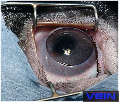
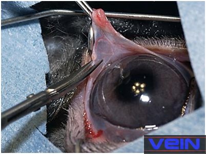
Akira Yamanouchi
Pre-surgical
preparation for the removal of the membrane growing over the cornea
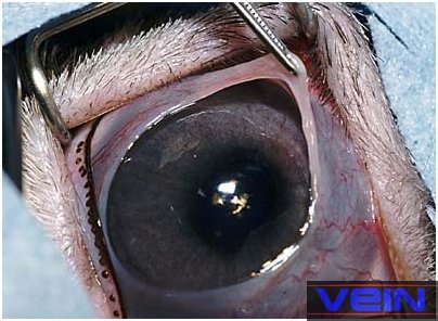
Akira Yamanouchi
The membrane is removed, by cutting it in 4 to 6 segments, from the
edge back to the conjunctival bulbar tissue. Once placed in it normal
position, the membrane is sutured to the eyelid, the sclera or the fornix
conjunctivae.
Sutures can be removed after 3 weeks, or left in place.
Acknowledgement
My gratitude goes to
Akira Yamanouchi (Veterinary
Exotic Information Network, Japan), for the
permission to use his pictures.
Further
information
Arnbjer, J.
Pseudopterygium in a pygmy rabbit. Vet. Med. Small Anim.
Clin. 74,737-738 (1979).
Bourne D. Aberrant
conjunctival overgrowth in rabbits.
http://wildlife1.wildlifeinformation.org/S/00dis/Miscellaneous/AbConjunctOvergrowthRabbit.html
Delaney, K.H.
Diagnostic exercise: Apparent corneal occlusion in a New Zealand white
rabbit. Contemp. Top. Lab. Anim. Sci. 34,76-77 (1995).
DuPont, C., Carrier,
M. & Gauvin, J. Bilateral precorneal membranous occlusion in a dwarf
rabbit. J. Small Exotic Anim. Med. 3,41-44 (1995).
Fehr, M. Eye anomalies
in dwarf rabbits. [German].
Kleintierpraxis 29, 129-130, 132 (1984).
Matros, L.E., Ansari,
M.M. & Van Pelt, C.S. Eye anomaly in a dwarf rabbit. Avian Exotic
Pract. 3,13-14 (1986).
Roze, M., Ridings, B.
& Lagadic, M. Comparative morphology of epicorneal conjunctival membranes
in rabbits and human pterygium. Vet. Ophthalmol. 4,171-174 (2001).
Schoofs, S. &
Hanssen, P. Epicorneal conjunctival membrane in the rabbit: a clinical case
and surgical therapy. Vlaams Diergeneeskundig Tijdschrift 67,344-346 (1998).
Wagner, F., Heider,
H.J., Gorig, C. & Fehr, M. Ophthalmic diseases in dwarf rabbits. Part 1:
eye examination, anatomy, diseases of the eyelids, the conjunctiva and of the
nasolacrimal duct. [German]. Tierarztl. Prax. 26,205-210
(1998).
|
e-mail: info@medirabbit.com
