![]()
Corneal
lipidosis or lipid deposit in the cornea of rabbits
Esther van Praag, Ph.D.
|
|
MediRabbit.com is
funded solely by the generosity of donors. Every
donation, no matter what the size, is appreciated and will aid in the
continuing research of medical care and health of rabbits. Thank you |

Warning: this file
contains pictures that may be distressing for people.
Corneal lipidosis - also called corneal
dystrophy or lipid keratopathy - is a condition where excess lipids (usually
cholesterol esters) or minerals (calcium) are deposited under the surface of
the cornea. The infiltration usually starts at the edge of the cornea and can
be observed in the anterior stroma, the epithelial basement membrane and the
epithelium.
Corneal lipidosis is not associated to a disease; it is not breed or
gender dependent.
Etiology
A lipid rich diet and/or trauma are the main causes for lipid
deposits into the cornea. Congenital factors cannot be ruled out.
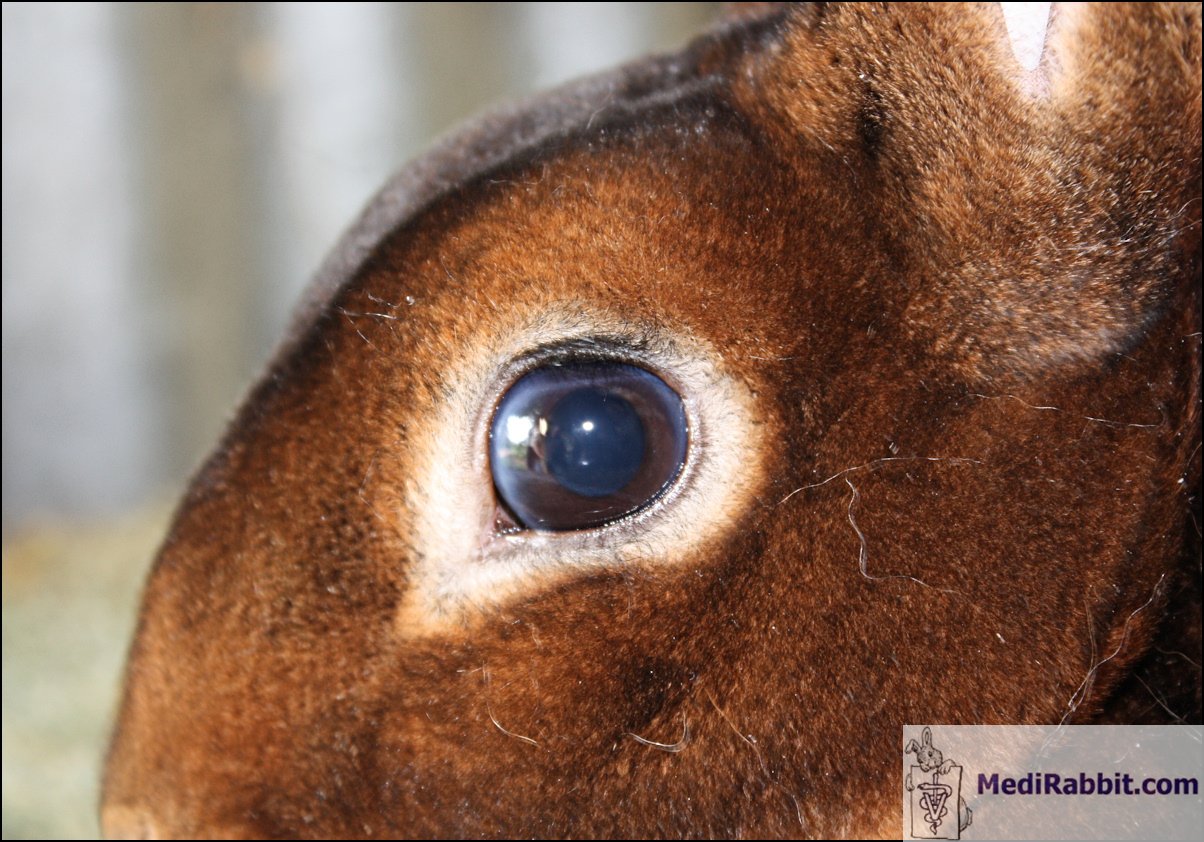
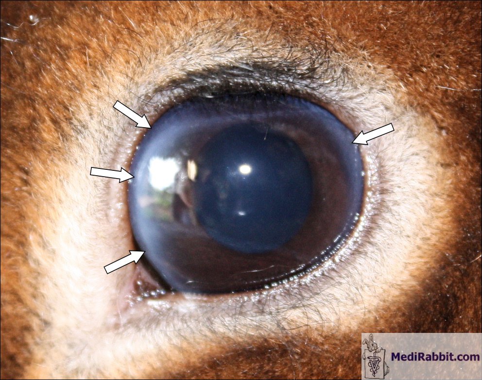
Michel Gruaz Rabbit
presenting a circle opacity at the periphery of the
cornea, caracteristic sign of lipid depsits. The disease is called lipid keratopathy or lipidosis f the cornea.
Clinical signs
It is based on
a complete ophthalmic examination and a discussion with the owner about the
food fed to the rabbit.
Both eyes are usually affected (bilateral) but not necessarily to the
same degree. Unilateral lipidosis has rarely been reported. The fat deposits,
which usually start near the third eyelid, can be opaque, raised, subtle and
pale, bright white, silver or grey colored areas. Vascularization is observed
in the affected part of the cornea. While the cornea is mainly affected, fat
deposits have also been noted in the lens, iris and ciliary body of a Dutch
rabbit. Often it is accompanied by macrophage invasion. An inflammatory
process has been observed, but does not always seem to be present.
Unlike in dogs,
corneal lipidosis is associated to gradual loss of vision in rabbits. If the
deposit is severe, it can lead to ulceration of the cornea.
There is no pain associated to this condition.
Diagnostic
and differentiel
A blood test helps confirm the high level of lipids and
cholesterol in the blood. The ratio of LDL and HDL corresponds to that
present in the eye.
A complete eye examination is necessary to confirm the
diagnosis.
A discussion of the rabbit’s diet is necessary and
corrections must be immediately implemented.
Corneal lipidosis should be
differentiated from:
- Retinal lesions, when loss of vision is observed;
-
Neoplasia;
-
Uveitis;
- Cerebral lesions, even when these are generally
accompanied by further neurologic signs, while the persistant
physiological constriction of the pupilla (photomotor reflex or pupillar
reflex) is not affected.
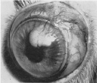
https://circ.ahajournals.org/cgi/reprint/18/4/519
Lipid plaque in hypercholesteremic rabbit cornea
occurring at site of vascularity. Cornea had been cauterized several times by heated probe.
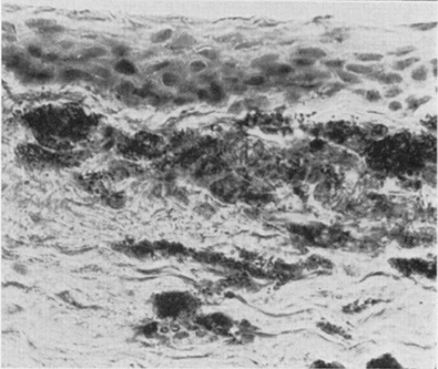
https://circ.ahajournals.org/cgi/reprint/18/4/519
Sections
of rabbit cornea in region of plaque stained with hematoxylin-Sudan.
Noteworthy is abundance of intracellular globular lipid and relatively slight
amount of granular sudanophilia.
Treatment
There is no medical therapy available, other than bring modification
to the diet. Fatty food and milk-based products (cheese, butter, and yogurt)
should be discontinued.
It is not known if superficial keratotomy can help.
Acknowledgement
Many thanks to Michel Gruaz (Switzerland)
for his great pictures of lipid accumulation in this Rex rabbit and his
permission to use on this page.
Further information
Fallon MT, Reinhard MK,
DaRif CA, Schoeb TR. Diagnostic exercise: eye lesions in a rabbit. Lab Anim Sci. 1988;
38:612-3.
Garibaldi BA, Goad ME.
Lipid keratopathy in the Watanabe (WHHL) rabbit. Vet
Pathol. 1988; 25:173-4.
Gwin RM, Gelatt KN.
Bilateral ocular lipidosis in a cottontail rabbit fed an all-milk diet. J Am Vet Med Assoc. 1977; 171:887-9.
Hillyer E.V., Quesenberry K.E.,
Ferrets, Rabbits, and Rodents: Clinical Medicine and Surgery, New York: WB
Saunders Co., 1997, p. 339-345.
Sebesteny A, Sheraidah
GA, Trevan DJ, Alexander RA, Ahmed AI. Lipid keratopathy and atheromatosis in
an SPF laboratory rabbit colony attributable to diet. Lab Anim. 1985;
19:180-8.
Stock EL, Mendelsohn
AD, Lo GG, Ghosh S, O'Grady RB. Lipid keratopathy in rabbits. An animal model
system. Arch Ophthalmol. 1985 May;103(5):726-30.
|
e-mail: info@medirabbit.com
