![]()
Prolapse of the
Harderian gland or “cherry eye”
Esther van Praag, Ph.D.
|
MediRabbit.com is funded solely by the
generosity of donors.
Every donation, no matter what
the size, is appreciated and will aid in the continuing research of medical care
and health of rabbits.
Thank you
|

Warning: this file contains
pictures that may be distressing for people.
This sebaceous gland has been given several names: Harderian gland,
Harder’s gland, nictitans gland, deep nictitating
membrane, or the medical terms: glandula palpebrae tertiae superficialis or profunda.
The gland was first discovered in the red deer by the Swiss physician
Harder (1694). It was subsequently found to be present in amphibians,
reptiles, birds and mammals that possess a third eyelid.
The Harderian gland is located within the eye orbit, at the nasal base of the third eyelid. In rabbits, it is
composed of 2 lobes:
• A dorsal white lobe;
• A ventral pink lobe.
The white lobe is small, on the contrary of the pink lobe. They cannot
be differentiated from each other during a histological analysis, in spite of
their different color. Intact male rabbits have a particular large Harderian
gland, which increases further in size during the breeding season.
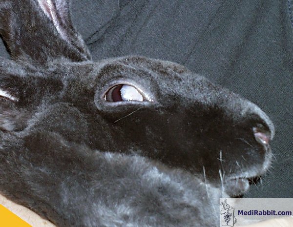
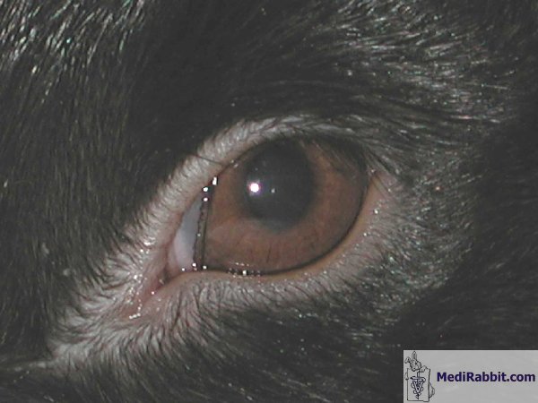
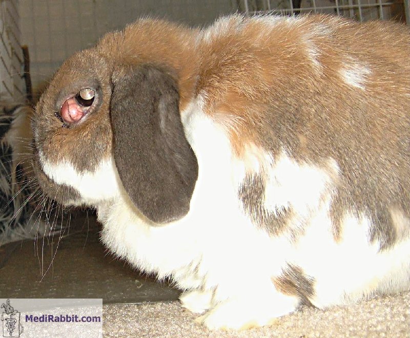
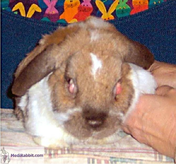
A swelling of the third eyelid occurs when the ventral pink lobe of
the Harderian gland prolapses. The etiology of the prolapse is unknown. A
weakness of the connective tissue around the gland is suspected. The gland
starts to move, and becomes irritated. Irritation leads to swelling and
sometimes discharge. The third eyelid can become bloody and ulcerated, and
develops a follicular conjunctivitis.
Diagnosis
The clinical signs should be
sufficient for a proper diagnosis. The condition of prolapsed Harderian gland
must be differentiated from a retrobulbar fat prolapse or retrobulbar
malignant B-cell lymphoma. Malignant B-cell lymphoma was of the Harder’s
gland was discovered in a 22 years old rabbit. The retrobulbar mass caused
unilateral exophthalmoses. After euthanasia, a necropsy showed the presence
of enlarged mesenteric lymph nodes. The cecum and the kidneys were also
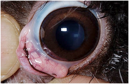
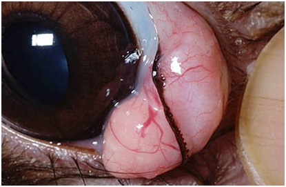
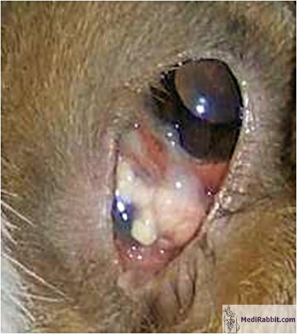
affected. The color and appearance of the prolapsed tissue should be
indicative: pink and lobular for the prolapsed Harderian gland, white for the
retrobulbar fat. The later condition is sometimes observed in obese rabbits.
Treatment
In the past years, the surgical procedure includes the surgical
removal of the gland. This is a difficult step in rabbits, as the gland is
located around the orbital venous sinus. It has, furthermore, been observed
that removal of the Harderian gland led to dry eyes in other animal’s
species.
Nowadays, the treatment of a rabbit suffering from a prolapsed
Harderian gland is similar as for dogs. The prolapsed gland is pushed back in
its pocket, in a slightly deeper position. Janssens
and Simoens describe the procedure the following
way: “a conjunctival incision is made dorsal to the prolapsed gland and an
anchoring suture is placed through the periosteum (dense fibrous connective
tissue surrounding bones) of the orbital rim. A horizontal bite is taken
through the gland dorsally and the anchoring suture passes ventrally through
the gland to exit through the conjunctival incision.”
The surgical procedure does not necessarily need a full anesthesia,
good sedation and local anesthesia is possible when the general health of the
rabbit is poor.
Acknowledgement
Thanks are due to Kim Chilson and to Akira
Yamanouchi (Veterinary Exotic Information Network, https://vein.ne.jp/), for
the permission to use their pictures.
Further information
Flecknell P., editor
Gloucester, BSAVA Manual of Rabbit Medicine and Surgery, UK: British Small
Animal Veterinary Association2000.
Hillyer E.V. and Quesenberry K.E., Ferrets, Rabbits, and Rodents: Clinical
Medicine and Surgery, New York: WB Saunders Co.1997.
Janssens G, Simoens P, Muylle S, Lauwers H. Bilateral prolapse of the deep gland of the
third eyelid in a rabbit: diagnosis and treatment. Lab Anim
Sci. 1999; 49(1):105-9.
Manning P.J., Ringler D.H., Newcomer C.E., The
Biology of the Laboratory Rabbit, New York: Academic Press1994.
Donnelly TM. Pink mass
on the dorsomedial aspect of a rabbit's eye: cherry eye or prolapse of the
deep gland of the nictating membrane. Lab Anim (NY). 2002;
31(2):23-4.
Richardson V.,
Rabbits: Health, Husbandry and Disease, Blackwell Science Inc
2000.
Volopich S, Gruber A, Hassan J, Hittmair
KM, Schwendenwein I, Nell B. Malignant B-cell
lymphoma of the Harder's gland in a rabbit. Vet Ophthalmol.
2005 Jul-Aug;8(4):259-63.
|
||||||||||
e-mail: info@medirabbit.com
