![]()
Cataract in
rabbits
Esther van Praag, Ph.D.
|
|
MediRabbit.com is
funded solely by the generosity of donors. Every
donation, no matter what the size, is appreciated and will aid in the
continuing research of medical care and health of rabbits. Thank you |

Warning: this file
contains pictures that may be distressing for people.
The word cataract comes from the Latin “cataracta”, or from
the Greek katarraktēs, meaning waterfall. It reminds the progressive
increase in opacity of the eye lens, which is sometimes referred to as
“looking though a waterfall”. Consequently, a reduced amount of light passes
through the lens. The ability to focus and eyesight sharpness will decrease
with time. This is accompanied by a loss of sensitivity to contrast.
The ability to see objects in bright light is reduced and an affected
rabbit will start to hop furniture or any object that is on its path. The
changes in the lens are due to the oxygen metabolism and the recycling of the
gluthatione protective molecule. Since the lens has no direct contact with
the blood circulation, its level of oxygen is the second lowest in the body,
after the nervous system and the adrenal cortex. Oxidative respiration, which
is carried out by the mitochondria organelles within the cells of the lens,
is sufficient to produce the ATP (form of stored energy in organisms)
necessary for the proper functioning of cells. This is accompanied by the
formation of free radicals and other oxidative molecules. These molecules are
neutralized by a smaller protein, gluthatione. While gluthatione is oxidized,
the free radicals are reduced and neutralized. The oxidized gluthatione will
move to the surface of the lens, where it will be reduced by the enzyme
gluthatione reductase, with the help of a co-enzyme derived from vitamin B3. The cycle
enables to regenerate the glutatione, so it can be used again. Vitamin C also
plays a protective role and its concentration within the lens is about 40
times higher than in the blood. Once the vitamin C has entered the lens
cells, it will also start to reduce free radicals and other oxidizing
molecules.
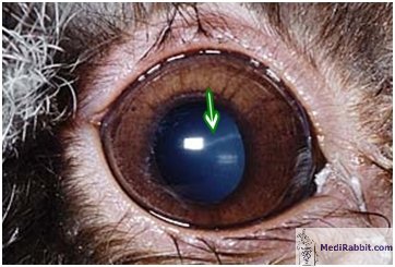
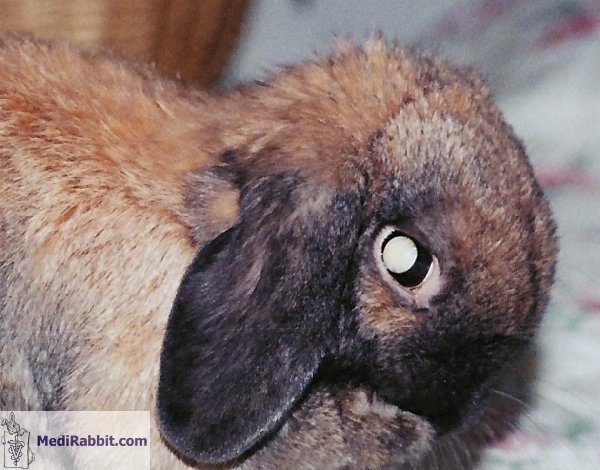
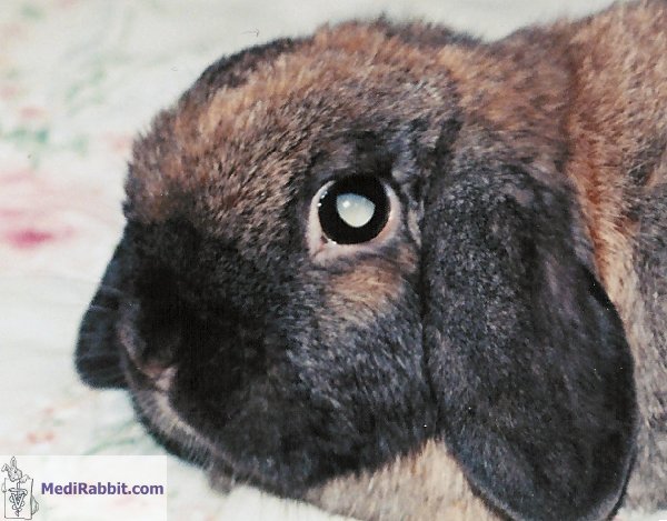
Cataracts
observed in mammals and human beings can be classified in three categories:
·
Nuclear cataract: it is
characterized by a degeneration of the proteins in the center of the lens
(nucleus) due to age. It is typically related to increased levels of oxidized
(dangerous) gluthatione in the lens. Possibly the movement of the later is
slowed down due to age or to a disbalance between proteins and lipid
oxidation. The lens becomes
white and later brown.
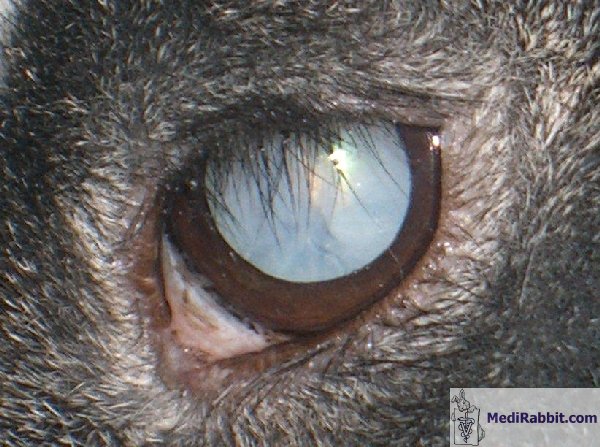
§ Cortical cataract: it is related to the disruption of the lens at
the periphery and spreads towards the center. It is typically related to a
decrease in the level of gluthatione, accompanied by the excessive
destruction of proteins, damage of the fiber plasma membrane or disruption of
the calcium homeostasis (maintained relatively constant state within the
body).
§ Posterior subcapsular cataract: it is characterized
by the development of clusters of swollen cells in the back of the lens.
Several clusters can develop, independently from each other. This type of
cataract is rare and typically stress induced (e.g. UV) or due to medication
(corticosteroids).
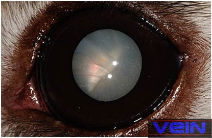
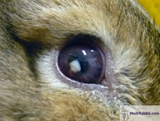
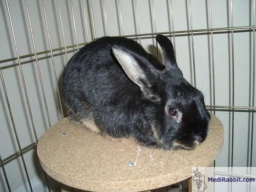
In rabbits,
there is a fourth cause for cataract and lens rupture, related to protozoal
parasite Encephalitozoon cuniculi.
Causes
The appearance of cataract is generally related to age. Further
contributing factors are heredity, nutrition, medication, exposure to sun
light, presence of the protozoal parasite Encephalitozoon cuniculi,
head-trauma, or a diet poor in caretonoids. The incidence and the causes of
cataract in rabbit is not well known. On the contrary to other animals,
cataract development is not related to diabetes, a metabolic disorder that is
very rare seen in rabbit.
Increased oxidative stress,
due to the presence of free radicals, a breakdown of the protective
mechanism, or a decreased gluthatione cycle, lead to an accumulation of
hydrogen peroxide in the aqueous humor of the eye. Although gluthatione will
reduce the peroxide, the energy-producing metabolism will be destroyed on the
long term, enabling the diffusion of sodium into the lens. Osmolality
(natural tendency to maintain water balance) will lead to edema (accumulation
of water) in the lens. The proteins inside the lens oxidize, become opaque
and insoluble (similar process as heat induced denaturation of ovalbumin and
egg white proteins).
The free radicals attack the lipids present in the membrane, which
leads to a shrinkage or swelling of the lens capsule. These changes of
pressure inside the lens break the lens fiber membranes and the space will be
filled with water and waste.
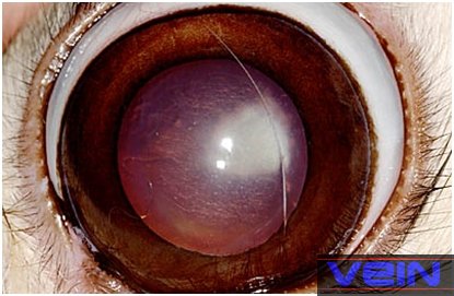
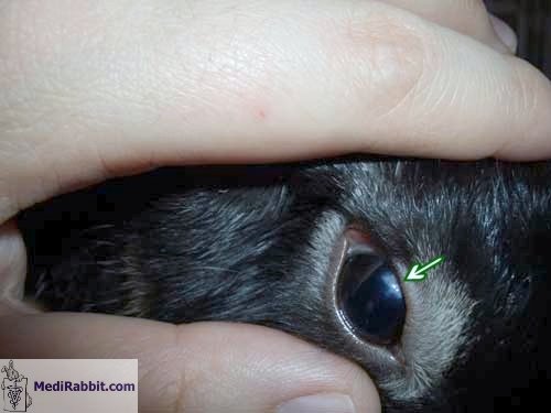
Diagnosis
A comprehensive diagnosis enables to diagnose a
cataract and monitor its development with time. Rarely, lens rupture is
observed.
Treatment
The treatment of choice for cataract is surgical removal, using the
phacofragmentation of the lens techniques, without replacement of the lens.
Indeed, regeneration of the lens has been observed in numerous rabbits. If
this will not be the case, the rabbit will nevertheless be able to discern light
and differentiate shapes.
If the cause of cataract relates to the parasite E.
cuniculi, living in the nervous system of rabbits, the treatment
includes the administration of fenbendazole (20 mg/kg, q 24 h., during a
month) during 28 days. Longer treatments must be avoided as onset of
secondary effects such as bone marrow depression has been observed in
rabbits. The use of albendazole should be avoided in rabbits. It has lead to
sudden death in healthy and/or young rabbits, immediately after
administration of the drug (private communication, Prof. P. Deplazes, DVM,
Faculty of Veterinary Medicine, University of Zurich, Switzerland)
If uveitis is present and the lens cannot be removed surgically, the
use of a topical NSAID or non-NSAID medication (e.g. prednisolone acetate 1%)
is necessary.
Cataract may cause pain. If this is observed, the use of analgesics
is recommended. If treatment does not bring relief or improvement, eye
surgery or enucleation may help the rabbit.
Acknowledgement
My gratitude goes to
Amy Carpenter (USA), to Susan L. (USA), to Lisa Hutcheon (USA), to Christine
Goodhand, to Melanie Kuenzel and Heather Bechtel (USA), to Sandy Minshull
(Canada) and to Akira Yamanouchi (Veterinary
Exotic Information Network), for the permission
to use their pictures and/or their help. Many thanks also to the rabbits that
helped illustrate this page.
Further
information
Arnesen K, Nordstoga K. Ocular encephalitozoonosis (nosematosis) in blue foxes. Polyarteritis nodosa and cataract. Acta Ophthalmol (Copenh). 1977; 55:
641-51.
Ashton N, Cook C, Clegg F. Encephalitozoonosis
(nosematosis) causing bilateral cataract in a rabbit.
Br J Ophthalmol. 1976; 60: 618-31.
Felchle LM, Sigler RL. Phacoemulsification for the management of Encephalitozoon
cuniculi-induced phacoclastic
uveitis in a rabbit. Vet Ophthalmol. 2002;
5: 211-5.
Flecknell P., editor
Gloucester, BSAVA Manual of Rabbit Medicine and Surgery, UK: British Small
Animal Veterinary Association2000.
Gelatt KN. Congenital
cataracts in a litter of rabbits. J Am Vet Med Assoc. 1975; 167:598-9.
Giordano C, Weigt A,
Vercelli A, Rondena M, Grilli
G, Giudice C. Immunohistochemical
identification of Encephalitozoon cuniculi in phacoclastic
uveitis in four rabbits. Vet Ophthalmol.
2005; 8: 271-5.
Gwon A, Gruber LJ,
Mantras C. Restoring lens capsule integrity enhances lens regeneration in New
Zealand albino rabbits and cats. J Cataract Refract Surg. 1993; 19: 735-46.
Gwon A, Gruber L,
Mantras C, Cunanan C. Lens regeneration in New Zealand albino rabbits after
endocapsular cataract extraction. Invest Ophthalmol Vis Sci. 1993; 34:
2124-9.
Harcourt-Brown F.,
Textbook of Rabbit Medicine, Oxford, UK: Butterworth-Heinemann, 2001.
Quesenberry K.E.,
Carpenter J.W., Quesenberry P., Ferrets, Rabbits and Rodents: Clinical
Medicine and Surgery Includes Sugar Gliders and Hedgehogs. Elsevier Health, 2004.
11. Weisse I, Niggeschulze A, Stotzer H. Spontaneous
congenital cataracts in rats, mice, and rabbits. Arch Toxicol. 1974;
32:199-207.
|
|||||||||||||||||||||

e-mail: info@medirabbit.com
