![]()
Causes for corneal ulcers in rabbits
|
MediRabbit.com is funded solely by the
generosity of donors.
Every donation, no matter what
the size, is appreciated and will aid in the continuing research of medical care
and health of rabbits.
Thank you
|

|
Akira Yamanouchi Rabbit
eye with a corneal ulcer as seen with normal light |
Akira Yamanouchi Same
eye, seen with black light (Wood's light), after application of the fluorescein dye |
|
|
|
|
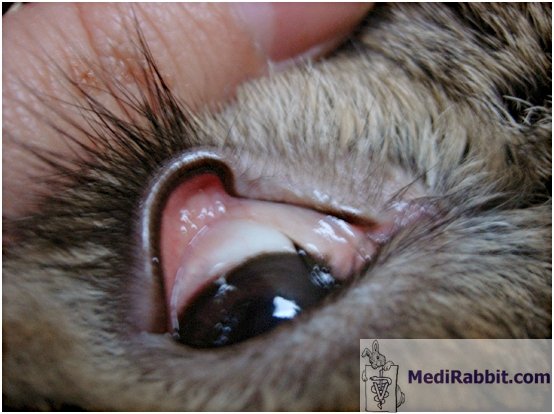
Ivy Two year old French lop rabbit
suffering from entropion (red arrow) and eye lashes falling on the cornea,
accompanied by secondary dacryocystitis (green arrow). This condition can
cause damage to the cornea and can be corrected surgically. |
|


|
Traumatic
causes |
Bedding,
e.g., hay or straw dust in the eye, bars of cage. |
|
Fighting,
scratch by a cat’s nail |
|
|
Excessive
rubbing, due to the presence of a foreign body, irritation, |
|
|
Self-trauma,
secondary to conjunctivitis, boredom, irritation, pain |
|
Viral
causes |
Myxoma virus
|
|
Secondary to bacterial causes |
Presence of
bacteria
|
|
Blepharoconjunctivitis, or a chronic inflammatory infection
of the eyelash and oil glands of the eyelids, accompanied by the inflammation
of the membrane covering the surface of the eyeball. This disease can relate
to the presence of bacteria or viruses (e.g., Myxoma virus). |
|
|
Chronic
inflammation of the nasolachrymal duct, in that case, the ulcers are
typically ventral or superficial. |
|
Eyelid disease |
Entropion with
the edge of the eyelid that turns inward toward the eye.
|
|
Distichiasis
with abnormal growth of an eyelash from the meibomian glands along the eyelid
margin. |
|
|
Trichiasis, an acquired condition in which the
eyelashes are misdirected and grow inwards towards the globe. |
|
|
Crenated eyelid |
|
“Mechanical”
causes |
Dry eyes due to
a hindered production of tears.
|
|
Special eye conditions |
Exophthalmia, causes by a retrobulbar mass (B-cell lymphoma), an
abscess
|
Ulcer development, secondary to glaucoma.
|
|
Diet |
Mineral or vitamin
deficiencies, e.g., vitamin A deficiency
|
|
Surgical
causes |
Exposure keratopathy, due to anesthesia.
|
A big thank you to Ivy (Serbia) and
Akira Yamanouchi (Japan, Veterinary Exotic Information Network, https://vein.ne.jp/)
for the permission to use their pictures.

e-mail: info@medirabbit.com
