![]()
Rabbit skull
radiology
Corby Holson
and David Martinez-Jimenez
|
|
MediRabbit.com is
funded solely by the generosity of donors. Every
donation, no matter what the size, is appreciated and will aid in the
continuing research of medical care and health of rabbits. Thank you |

|
Introduction to rabbit radiology Radiology in the domestic rabbit (Oryctolagus cuniculus) can be
challenging due to their size, and easily stressed nature. Small mammal
radiology requires a basic knowledge of anatomy and physiologic
characteristics. Therefore, three important factors require further attention
when taking radiographs in rabbits: 1.
Rabbits are prey species and therefore, they become
extremely agitated when exposed to unfamiliar situations. Manual restraint
can be difficult, and dangerous, for any length of time. Sedation
or anesthesia is usually required. 2. The relatively small size of
rabbits has the advantage of allowing whole-body radiographs and quick
examination of the entire patient. However, it may result in a loss of detail
due to the differences in size between different body areas. 3. Motion of the object during the
imaging sequence generally results in a blurring. Because motion artifact is
a common problem in rabbits, the minimum requirement for x-ray equipment is
the capacity to produce 300 mA in 1/120 (0.008) second. Settings and equipment The x-ray machine should be capable of working with 5.0 to 7.5 mAs
exposures, and have a range of 40 to 100 KVp, which is adjusted in 1 to 2 KVp
increments. Often when radiographing rabbits, mammography films and cassettes
are utilized (Mamoray® cassette, Mamoray HDS, AGFA Corporation, Greenville,
SC). This system provides ultra fine detail in smaller species. Positioning Radiography in rabbits, and other small mammals, can be challenging as
they are highly stressful, resistant to restraint and have a small body size
in relation to their short extremities. Visual anatomic landmarks are
preferred over palpated anatomic landmarks because their thick subcutaneous
adipose layers and dense hair coats make it difficult to palpate accurately.
Correct symmetry and stabilization of the patient can be accomplished by
using radiolucent materials such as foam and tape for support. Skull radiography Rabbit bones are delicate in comparison to that of other domestic
companion animals. For example, the skeleton of a rabbit represents only 7-8%
of body weight but is 12-13% of body weight in a cat. Paradoxically, the
muscles of rabbits are extremely strong and powerful which can lead to
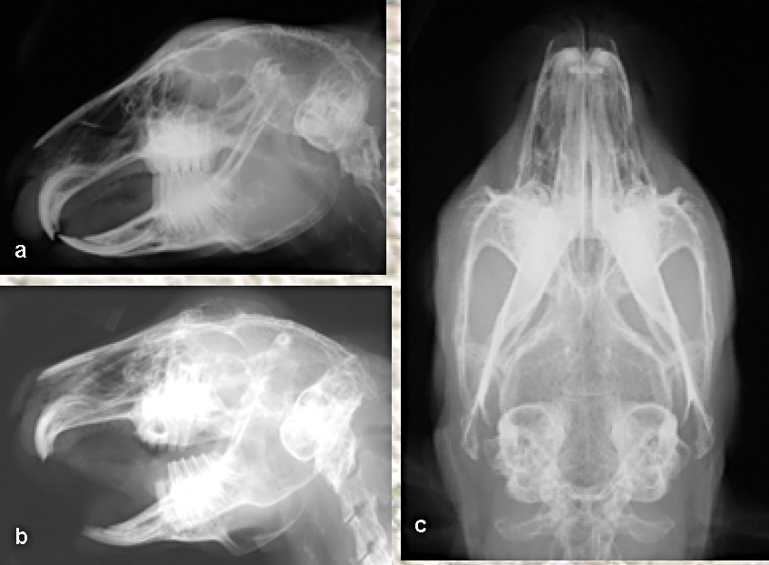
fractures or luxations if held improperly. Skull radiography can provide useful information about the nares,
sinuses, middle ears, teeth and surrounding bone. Radiographic views should
include lateral and dorsoventral views, as well as obliques when necessary
(e.g. dental disease evaluation). Oblique radiographs projections require
rotations at 30 degrees angles (described by the point of entrance of the
x-ray beam to the point of exit). Magnification radiography is
commonly used in exotic patients when higher definition, detailed images are
necessitated. It requires the use of an ultra-small focal spot x-ray tube and
an increased object-film distance as compared to standard radiographs.
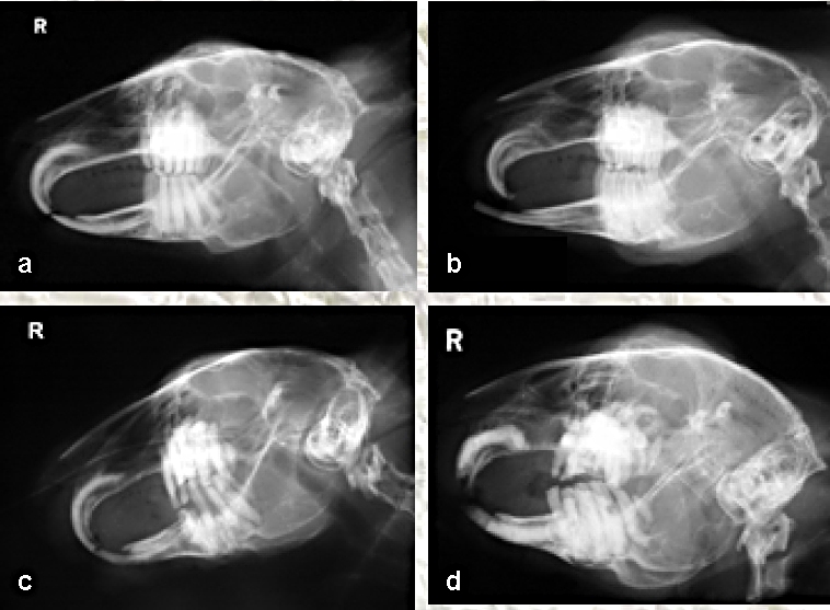
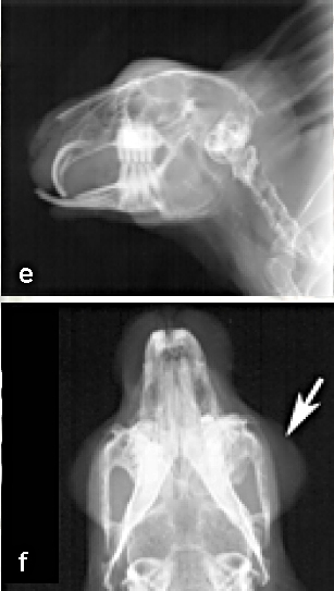
Pathological radiography associated to the rabbit skull Skull radiography is an extremely valuable tool in
rabbit medicine, not only because it helps localize and diagnosis specific
anatomical disease, but it also serves as an important prognostic
determinant. For instance, an increased density in tympanic bullae, nares, or
sinuses may indicate infection. On the other hand, decreased density may
occur in advanced cases of infection or neoplasia.
Degree of dental disease and prognosis can be also
determined by radiographic evaluation. Facial abscesses and osteomyelitis of
the maxilla or mandible in lagomorphs are often related to dental disease and
bacterial infection. The precise etiology of dental disease in the pet rabbit
is still unknown and is likely to be multifactorial including primary
(inherit) and secondary causes (diet, metabolic, traumatic and infectious
components).
Radiography is an essential diagnostic tool that can
help localize both bone and soft tissue lesions before attempting surgical
correction. Radiographs can also accurately grade dental disease from 1
(normal) to 5 (severe dental disease, including oral abscesses). Conclusion Radiology is an essential tool for any practitioner
working with rabbits. Due to their stressful temperament and keen ability to
hide disease, thorough and careful examination including radiographs is
warranted. Rabbits are particularly predisposed to dental disease, and skull
radiographs can detect lesions unlikely to be observed during physical
examination. Radiographs also serve as an important tool for grading dental
disease and are valuable in providing accurate prognosis to clients. References Harcourt-Brown, F. M. (1997).
"Diagnosis, treatment and prognosis of dental disease in pet
rabbits." In Practice 19: 407-421. Lobprise, H. B. and R. B. Wiggs (1991).
"Dental and oral disease in Lagomorphs." J Vet Dent 8(2): 11-7. Silverman, S. and L. A. Tell (2005).
“Radiology equipment and positioning techniques”. In: Radiology of rodents, rabbits, and ferrets: an atlas of normal anatomy and
positioning. Elsevier Saunders, St Louis. Pp. 1-8. Stefanacci, J. D. and H. L. Hoefer
(2004). “Radiology and ultrasound”. In Ferrets, rabbits, and rodents.
Clinical medicine and surgery.” Second edition. K. Quesenberry and
J. W. Carpenter (Eds.). Elsevier Saunders, St. Louis. Pp. 395-396. Acknowledgement The authors would like to thank
Dr. Hernandez-Divers and Dr. Wilson for their help and support, as well as
for some of the pictures provided. |
e-mail: info@medirabbit.com
