![]()
Myxoid sarcoma
or myxosarcoma in rabbits
Esther van
Praag, Ph.D.
|
|
MediRabbit.com is
funded solely by the generosity of donors. Every
donation, no matter what the size, is appreciated and will aid in the
continuing research of medical care and health of rabbits. Thank you |

Warning: this file
contains pictures that may be distressing to some persons
|
The word “sarcoma” comes from the Greek and means
“fleshy growth”. Sarcoma is nowadays used to describe relatively rare a group
of malignant tumors that involve the connective tissue. Although sarcomas are
well-recognized tumors, their characteristics lead to confusion. Indeed, some
type of sarcoma may present a combination of features of various different types
of sarcoma. This lead to the widely accepted conclusion that the neoplastic
development of a primitive mesenchymal cell can lead into different
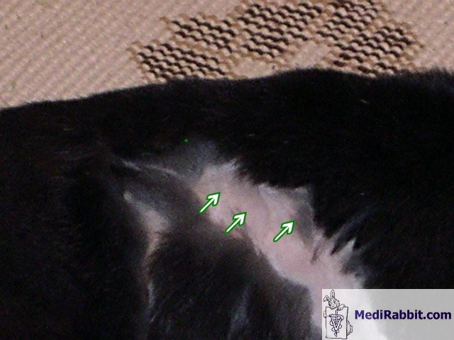
direction, thus different types of sarcoma. The tumors are found is all parts of the body:
forelimbs, hind limbs, head, neck, shoulder, chest, abdomen or hip; as well
as in all types of tissues: muscle tissue, fat tissue, in the blood vessels,
in the tissue surrounding joints, and in tendons. Four
principles apply for sarcoma tumors: • Location: a superficially located tumor is
less likely to be malignant than a deeper located tumor. • Size: bigger tumors are more likely to
become malignant than small tumors. • Growth: rapid growth tumors are more
likely to be malignant than slow growing ones. • Vascularization: malignant tumors are rich
in blood vessels, whereas benign tumors are avascular or poorly vascularized. Sarcoma
tumors are locally invasive into the surrounding tissues. Although their
metastatic rate is low, they can metastase through
the bloodstream to other organs. Myxosarcoma is defined has a fibrosarcoma
rich in connective tissue proteoglycans (mucins). This kind of tumor is
therefore also sometimes called myxoid fibrosarcoma. In veterinary medicine, the term “myxosarcoma” remains, however, a general term for
otherwise specific sarcoma tumors like myxofibrosarcoma, liposarcomas
or malignant fibrous histiocytomas. A virus called "malignant rabbit fibroma
virus" has been isolated in rabbits, which can lead to fibrosarcoma, and perchance to fibrosarcoma
derived myxosarcoma. See: “Fibrosarcoma in rabbit” Diagnosis and histology
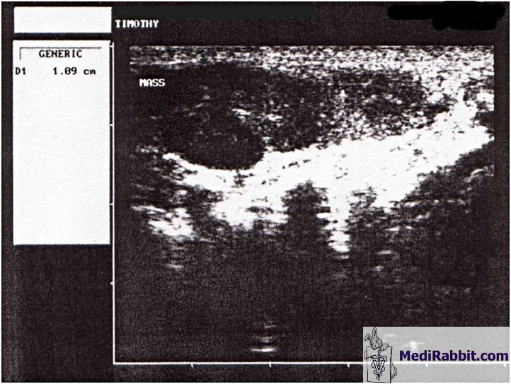
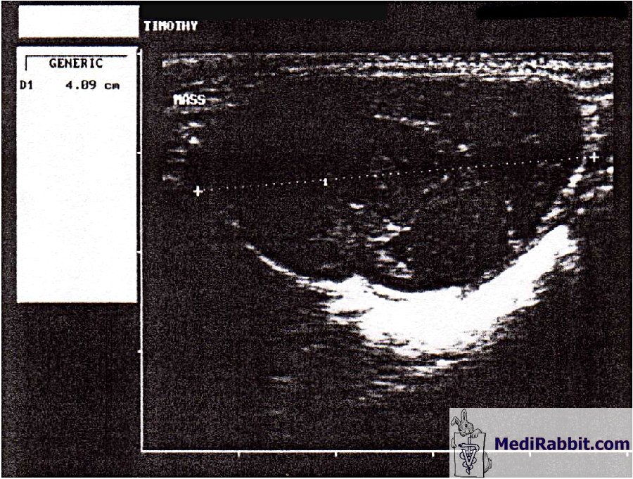
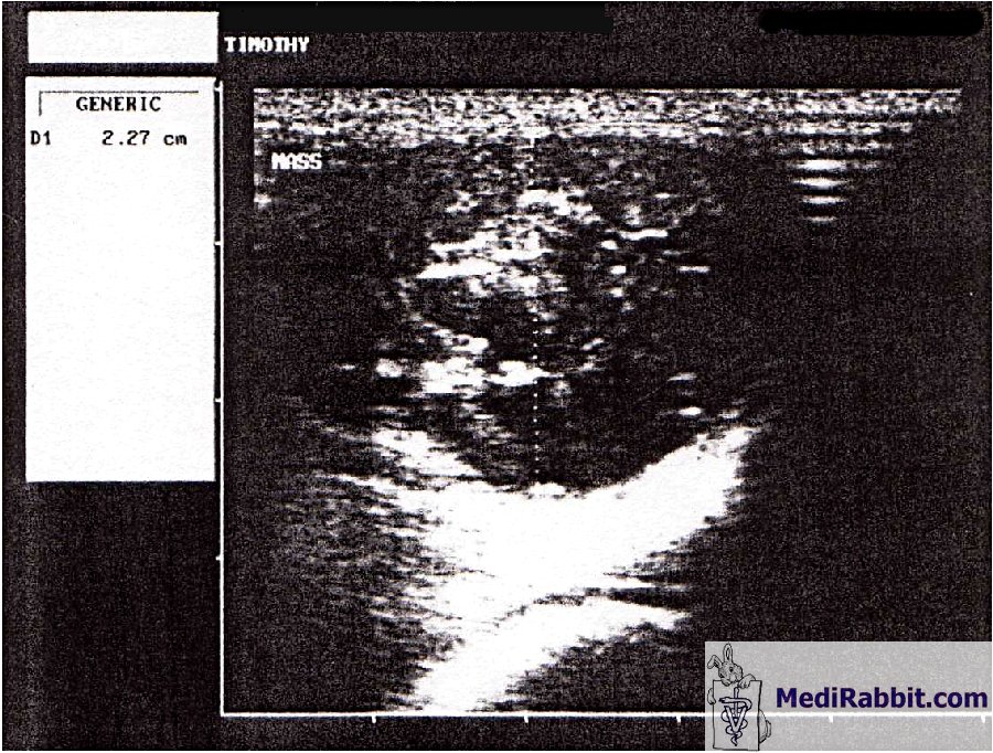
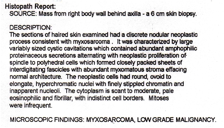
The nature of the
mass and the presence of metastases must be determined, by means of
X-ray, ultrasound, and a biopsy.
Myxosarcoma differentiate between
low-grade, intermediate and high-grade lesions. The tumors can be
small and (multi)nodular, or large. Their degree of malignancy is
generally low, but increases with successive regrowth (recurrence).
All have invasive properties
into surrounding tissues.
Typically, a tumor has a myxoid matrix containing
spindle- to polyhedral cells. The neoplastic cells can have various
shapes: round, ovoid or elongated; their nuclei is hyperchromatic.
Sheets of interdigitated cells, rich in myxoid
matrix can be present. Pseudolipoblasts
are commonly observed. Vascularization of the tumor is typical, with
curvilinear capillaries. A low-grade myxosarcoma must be differentiated from a benign myxoid lesion.
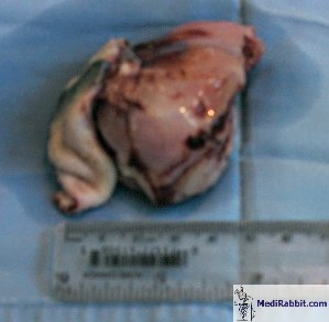
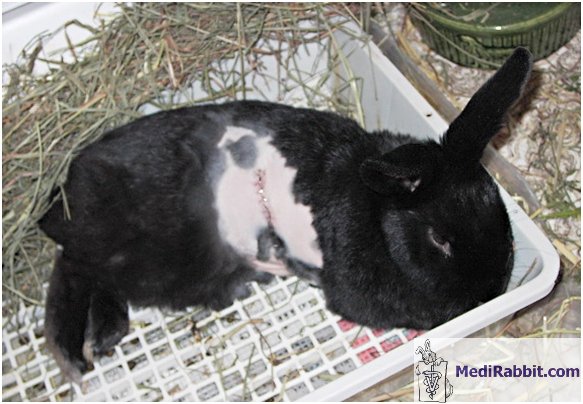
Treatment
The complete excision of the myxosarcoma lesions
is necessary. Indeed, a characteristic of myxosarcoma
and myxofibrosarcoma tumors is their high rate of recurrence (according
to species, up 70% within year of surgical removal). At recurrence,
these lesions gain a higher grade of aggressiveness and an increased
potential to become malignant. Prognosis is
guarded.
For detailed information on myxosarcoma in rabbits, see: “Skin
Diseases of Rabbits”, by E. van Praag, A. Maurer and T. Saarony, 408 pages, 2010.
Acknowledgements
All my gratitude
to Susan L. (USA), to Christine Harvey, DVM (USA), to E. Kufuor-Mensah,
DVM (USA) and to Dr. Taylor, DVM (USA) for kindly allowing the use
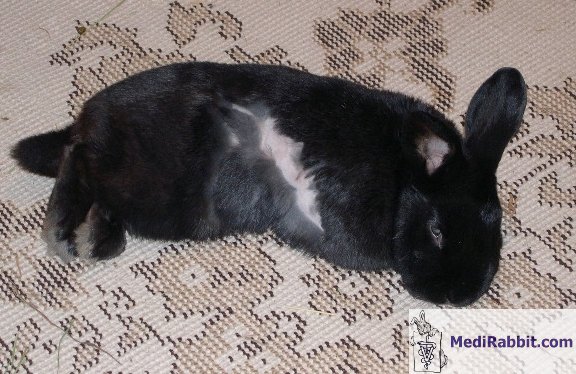
of illustrative material for this text. Thanks are also due to Timothy,
for his patience during picture-sessions. Further information
Flecknell P., editor Gloucester, BSAVA Manual of Rabbit Medicine
and Surgery, UK: British Small Animal Veterinary Association2000. Hillyer E.V. and Quesenberry K.E., Ferrets,
Rabbits, and Rodents: Clinical Medicine and Surgery, New York: WB
Saunders Co.1997. Janssens G, Simoens P, Muylle S, Lauwers H. Bilateral
prolapse of the deep gland of the third eyelid in a rabbit: diagnosis
and treatment. Lab Anim Sci. 1999; 49(1):105-9. Manning P.J., Ringler D.H., Newcomer C.E., The Biology of the Laboratory
Rabbit, New York: Academic Press1994. Richardson V., Rabbits:
Health, Husbandry and Disease, Blackwell Science Inc
2000. Schaff Z, Grimley PM, Michelitch J, Banfield WG. Spontaneous
myxosarcoma in a cottontail rabbit (Sylvilagus
floridanus): observation of tubular
structures in the endoplasmic reticulum of tumor cells. J Natl Cancer Inst.
1973; 51(1):293-7. Strayer DS, Sell S, Skaletsky E, Leibowitz JL. Immunologic
dysfunction during
viral oncogenesis. I. Nonspecific
immunosuppression caused by malignant rabbit fibroma virus. J Immunol. 1983; 131(5):2595-600. |