![]()
Deformed X and O limbs in rabbits
Esther van Praag Ph.D.
|
|
MediRabbit.com is funded solely by the generosity of
donors. Every donation, no matter what the
size, is appreciated and will aid in the continuing research of medical care
and health of rabbits.
Thank
you |

WARNING: this page may contain pictures that are distressing for some persons.
|
Rabbits
may exhibit limb deformities when viewed from the front or the side: ·
Acquired
or congenital angular deformation at the level of the limb joint; ·
Congenital
deformation and bowing of the long bones of limbs; ·
Laxity
or hypermobility of ligaments. The
etiopathology of angular limb deformation in rabbits is not well documented.
In other animals, including foals, there are several bone or muscle-related
causes. However, laxity (mobility) of the peri-articular structures and
ossification retardation of bones are often congenital in origin. Growth of
bones is influenced by a number of factors,
including genetic, hormonal (hypothyroidism), metabolic, and vascular or
electrical factors. Failure of one of these factors can lead to abnormal bone
growth and elongation of the longer bones in the limbs. In young growing
animals, asymmetrical mechanical pressure (e.g. the weight of the body or a
slippery floor, when the doe repeatedly removes hay and other materials from
the nest) on growing cartilages of long bones in the extremities can lead to
angular deformation of the joints, resulting in inward or outward deviation
of the limb. In such cases, the deformity will correct itself naturally once
the rabbit kit is placed in an environment without a slippery floor. In the
final stages of gestation, if a doe is not fed properly (for example, if she
is not getting the nutrients she needs, or if her diet is imbalanced), this
can also lead to limb deformation in her offspring. A
hereditary origin related to a single autosomal recessive gene appears to
cause bending of the upper limbs in some rabbit breeds, including Beveren
rabbits, Belgian giants, French Silver and Dutch
rabbits. The curvature of the limbs becomes apparent at the age of two to
three weeks. The upper limbs bend inwards, while the paws deviate laterally.
The deformity reaches its maximal stage at the age of 2 to 3 months. Even if
the cartilage problem corrects itself naturally by the age of two and a half
months, the deformity will be permanent, with inward curvature of the ulna
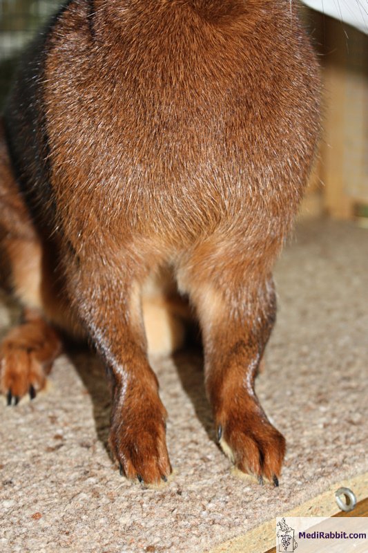
and radius (the forearm bones, located between the elbow and the front paw). Angular joint
deformation
Angular
joint deformities of the upper limbs are sometimes observed in rabbits. These
conditions are present before or soon after birth, before the closure of the
osseous growth plate. Lateral deviations are frequently observed in rabbits
and include: ·
Lateral,
valgus misalignment or X limb,
with lateral (outward) deviation of the limb distal to, or below the point of
deformity:
·
Medial,
in varus or in O, with a
lateral inward deviation of the limb distal to, or below the point of
deformity:
Osteo-articular
deformities are more prevalent in the upper limbs than the lower limbs of
rabbits. In some rare cases an upper may be affected by a double deformity at
the ulnar and carpal joints. It is usually present at birth or appears later
during growth, at around two to three weeks of age. Finally, angular
deformities can be accompanied by rotation of the limb. Faulty limb conformation
can have numerous causes
Ligament
laxity. It is usually present at the
newborn's birth and does not cause pain, but may also appear later in the
life of a rabbit. X-rays show that the morphology of the affected limbs
appears normal. This deformity generally resolves itself in foals. Rabbits
suffering from this problem may develop juvenile arthritis, which can result
in joint pain. In older
animals, deformity of the limbs in the sagittal plane can be indicative of
osteoarthritis. This syndrome is irreversible and painful, so it is essential
that the rabbit is administered analgesics. Treatment with glucosamine and chondroitin may also
help.
Cuboid
bones hypoplasia. The
cuboid bone is part of the tarsal bones. It rapidly undergoes a process of
ossification after birth. Thyroid dysfunction (hypothyroidism) is the
underlying cause of the lack of osseous development in foals, and hinders
normal ossification of the bones. A spontaneous correction is possible when
cartilage is still immature. Following this period, the deformity becomes
permanent. The deformed joint will undergo ossification and degenerative
changes. Furthermore, the onset of bone hypoplasia is irreversible. This
deformity carries with it an increased risk of partial luxation or fracture
of the limb. Deformation
of long bones. Growth
inequality between the medial and lateral parts of long bones results in
angular deformation of the limb, commonly in valgus or X. This deformity is
present at birth, but may also appear later, during the growing phase of the
young rabbit. In foals, the position in the uterus is thought to result in
abnormal compression of the growing cartilage during gestation. The trauma
results in unequal growth of the long bones. This type of deformity is not
associated with discomfort or pain. Preterm offspring and overweight. It has been observed that joint deformation is
also present in animals that are born prematurely, before the process of
calcification of bones takes place. Weight gain can result in angular
deformity of the joints in newborns. A similar problem has been observed in
single-born rabbit kits. At birth, they are larger in size, possess a robust
bone structure and exhibit accelerated growth, which is facilitated by the
does' adequate nourishment.
Clinical signs
A rabbit
suffering from deformed X or O-legs will have a normal appetite and be in
good health. The only clinical indications are an abnormal or difficult gait. Clinical examination
A rabbit
presenting articular deformities is examined from the front, the side and the back, at rest and during exercise on a
non-slippery surface. Palpation of the limb helps to evaluate the presence of
physical deformities, pain, swelling, and other potential issues. The
radiographic evaluation of limb bones and joints allows us to accurately
identify the origin of the problem, whether it is a primary congenital cause
or a secondary osteo-articular trauma. It is imperative that the front view
is perfect to facilitate the detection of lateral joint deformities. For the
purposes of evaluating sagittal deformation, both front and side views are
required. Treatment
If the
problem is discovered at a young age, a splint or rigid bandaging of the
affected limb may help correct the deformation. It is important to ensure
that young rabbits have sufficient space to move and exercise, as this is
beneficial for developing their muscles. There is
currently no treatment available for deformed limbs in rabbits. In cases
where limb function is significantly impeded or discomfort is being
experienced, amputation may be a beneficial course of action. In cases where
limb curvature and deformation are severe, the humane euthanasia should be
considered. Indeed, secondary complications of the skin may occur in areas
that support the body's weight. It is common to observe ulceration of the
skin, which is accompanied by pododermatitis and
is a source of significant discomfort and pain. Acknowledgements
I would like to express my sincere
gratitude to Michel Gruaz and Stefan Röthlisberger
(both from Switzerland) for their kind permission to use their pictures in
this article. Literature
references
Arendar GM, Milch
RA. Splay-leg - a recessively inherited form of femoral neck anteversion,
femoral shaft torsion and subluxation of the hip in the laboratory lop
rabbit: its possible relationship to factors involved in so-called
"congenital dislocation" of the hip. Clin Orthop. 1966;44:221-9. Barichard, T. Etude rétrospective du
traitement des déviations angulaires des poulains par des ondes de choc extracorporelles : 92 déviations. Thèse de médecine
vétérinaire. 2012. Wilson DG.
Les déformations angulaires des
membres chez le poulain: prise en charge et conséquences de l’angulation sur
la performance athlétique. la médecine vétérinaire des grands animaux. Rondes cliniques.
Vol 6, 2006. |
|||||||||||||||||||


